 Movie
Movie Controller

[English] 日本語
 Yorodumi
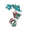
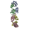
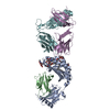
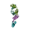
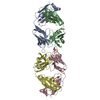
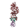
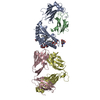
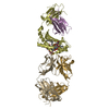
- PDB-1fj1: LYME DISEASE ANTIGEN OSPA IN COMPLEX WITH NEUTRALIZING ANTIBODY F... -
+ Open data

- Basic information
| Entry | Database: PDB / ID: 1fj1 | |||||||||
|---|---|---|---|---|---|---|---|---|---|---|
| Title | LYME DISEASE ANTIGEN OSPA IN COMPLEX WITH NEUTRALIZING ANTIBODY FAB LA-2 | |||||||||
 Components Components |
| |||||||||
 Keywords Keywords | IMMUNE SYSTEM / OSPA / LYME DISEASE / ANTIBODY FAB FRAGMENT / NEUTRALIZING EPITOPE | |||||||||
| Function / homology |  Function and homology information Function and homology informationInitial triggering of complement / Classical antibody-mediated complement activation / FCGR activation / Role of phospholipids in phagocytosis / Regulation of Complement cascade / phagocytosis, recognition / Regulation of actin dynamics for phagocytic cup formation / humoral immune response mediated by circulating immunoglobulin / positive regulation of type IIa hypersensitivity / positive regulation of type I hypersensitivity ...Initial triggering of complement / Classical antibody-mediated complement activation / FCGR activation / Role of phospholipids in phagocytosis / Regulation of Complement cascade / phagocytosis, recognition / Regulation of actin dynamics for phagocytic cup formation / humoral immune response mediated by circulating immunoglobulin / positive regulation of type IIa hypersensitivity / positive regulation of type I hypersensitivity / Fc-gamma receptor I complex binding / alpha-beta T cell receptor complex / immunoglobulin complex, circulating / phagocytosis, engulfment / immunoglobulin receptor binding / IgG immunoglobulin complex / immunoglobulin mediated immune response / complement activation, classical pathway / antigen binding / positive regulation of phagocytosis / B cell differentiation / cell outer membrane / positive regulation of immune response / antibacterial humoral response / adaptive immune response / cell surface / : / extracellular region / membrane / plasma membrane Similarity search - Function | |||||||||
| Biological species |  Borrelia burgdorferi (Lyme disease spirochete) Borrelia burgdorferi (Lyme disease spirochete) | |||||||||
| Method |  X-RAY DIFFRACTION / SYNCHROTRON / MOLECULAR REPLACEMENT / Resolution: 2.68 Å X-RAY DIFFRACTION / SYNCHROTRON / MOLECULAR REPLACEMENT / Resolution: 2.68 Å | |||||||||
 Authors Authors | Ding, W. / Lawson, C.L. | |||||||||
 Citation Citation | Journal: J.Mol.Biol. / Year: 2000 Title: Structural identification of a key protective B-cell epitope in Lyme disease antigen OspA. Authors: Ding, W. / Huang, X. / Yang, X. / Dunn, J.J. / Luft, B.J. / Koide, S. / Lawson, C.L. | |||||||||
| History |
|
- Structure visualization
| Structure viewer | Molecule: MolmilJmol/JSmol |
|---|
- Downloads & links
-Download
| PDBx/mmCIF format | 1fj1.cif.gz | 246.3 KB | Display | PDBx/mmCIF format |
|---|---|---|---|---|
| PDB format | pdb1fj1.ent.gz | 200.3 KB | Display | PDB format |
| PDBx/mmJSON format | 1fj1.json.gz | Tree view | PDBx/mmJSON format | |
| Others |  Other downloads Other downloads |
-Validation report
| Arichive directory | https://data.pdbj.org/pub/pdb/validation_reports/fj/1fj1ftp://data.pdbj.org/pub/pdb/validation_reports/fj/1fj1 | HTTPS FTP |
|---|
-Related structure data


-Links
 PDBj
PDBj


- Assembly
| Deposited unit | 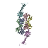
| ||||||||
|---|---|---|---|---|---|---|---|---|---|
| 1 | 
| ||||||||
| 2 | 
| ||||||||
| Unit cell |
| ||||||||
| Details | The biological assembly is an Fab/antigen complex composed of chains A, B, and F, or alternately C, D, and E |
-Components
| #1: Antibody | Mass: 23608.090 Da / Num. of mol.: 2 / Fragment: FAB FRAGMENT / Source method: isolated from a natural source Details: LA-2 HYBRIDOMA, GIFT OF M.M. SIMON, MAX PLANCK INSTUTUT, FREIBURG Source: (natural) #2: Antibody | Mass: 22629.268 Da / Num. of mol.: 2 / Fragment: FAB FRAGMENT / Source method: isolated from a natural source / Details: LA-2 HYBRIDOMA / Source: (natural) #3: Protein | Mass: 27682.195 Da / Num. of mol.: 2 Source method: isolated from a genetically manipulated source Source: (gene. exp.) Borrelia burgdorferi (Lyme disease spirochete)Strain: B31 / Plasmid: PET9C / Production host: #4: Water | ChemComp-HOH / |  Mass: 18.015 Da / Num. of mol.: 258 / Source method: isolated from a natural source / Formula: H2O Mass: 18.015 Da / Num. of mol.: 258 / Source method: isolated from a natural source / Formula: H2OHas protein modification | Y | |
|---|
-Experimental details
-Experiment
| Experiment | Method: X-RAY DIFFRACTION / Number of used crystals: 1 |
|---|
- Sample preparation
| Crystal | Density Matthews: 3.15 Å3/Da / Density % sol: 60.93 % | |||||||||||||||||||||||||
|---|---|---|---|---|---|---|---|---|---|---|---|---|---|---|---|---|---|---|---|---|---|---|---|---|---|---|
| Crystal grow | Temperature: 277 K / Method: vapor diffusion, hanging drop / pH: 6.15 Details: 100 mM sodium cacodylate, 100 mM sodium acetate, 10 % (w/v) PEG 3350, pH 6.15, VAPOR DIFFUSION, HANGING DROP, temperature 4K | |||||||||||||||||||||||||
| Crystal grow | *PLUS | |||||||||||||||||||||||||
| Components of the solutions | *PLUS
|
-Data collection
| Diffraction | Mean temperature: 100 K |
|---|---|
| Diffraction source | Source: SYNCHROTRON / Site: NSLS  / Beamline: X25 / Wavelength: 1.1 / Beamline: X25 / Wavelength: 1.1 |
| Detector | Type: BRANDEIS / Detector: CCD / Date: May 1, 1997 / Details: COLLIMATOR |
| Radiation | Monochromator: SI(111) / Protocol: SINGLE WAVELENGTH / Monochromatic (M) / Laue (L): M / Scattering type: x-ray |
| Radiation wavelength | Wavelength: 1.1 Å / Relative weight: 1 |
| Reflection | Resolution: 2.68→20 Å / Num. all: 51741 / Num. obs: 221681 / % possible obs: 97.8 % / Observed criterion σ(F): 0 / Observed criterion σ(I): 0 / Redundancy: 4.3 % / Biso Wilson estimate: 50.4 Å2 / Rmerge(I) obs: 0.048 / Net I/σ(I): 19.9 |
| Reflection shell | Resolution: 2.68→2.78 Å / Redundancy: 4 % / Rmerge(I) obs: 0.112 / Num. unique all: 5136 / % possible all: 98.4 |
| Reflection | *PLUS Num. obs: 51741 / Num. measured all: 221681 |
| Reflection shell | *PLUS % possible obs: 98.4 % / Num. unique obs: 5136 / Num. measured obs: 20967 |
- Processing
| Software |
| |||||||||||||||||||||||||
|---|---|---|---|---|---|---|---|---|---|---|---|---|---|---|---|---|---|---|---|---|---|---|---|---|---|---|
| Refinement | Method to determine structure: MOLECULAR REPLACEMENT Starting model: PDB ENTRY 1GAF, PDB ENTRY 1OSP Resolution: 2.68→19.83 Å / Rfactor Rfree error: 0.004 / Data cutoff high absF: 1428286.9 / Data cutoff high rms absF: 1428286.9 / Data cutoff low absF: 0 / Isotropic thermal model: GROUP / Cross valid method: THROUGHOUT / σ(F): 0 / σ(I): 0 / Stereochemistry target values: Engh & Huber
| |||||||||||||||||||||||||
| Solvent computation | Solvent model: FLAT MODEL / Bsol: 35.6764 Å2 / ksol: 0.318791 e/Å3 | |||||||||||||||||||||||||
| Displacement parameters | Biso mean: 52.8 Å2
| |||||||||||||||||||||||||
| Refine analyze |
| |||||||||||||||||||||||||
| Refinement step | Cycle: LAST / Resolution: 2.68→19.83 Å
| |||||||||||||||||||||||||
| Refine LS restraints |
| |||||||||||||||||||||||||
| Refine LS restraints NCS | NCS model details: CONSTRAINED | |||||||||||||||||||||||||
| LS refinement shell | Resolution: 2.68→2.85 Å / Rfactor Rfree error: 0.012 / Total num. of bins used: 6
| |||||||||||||||||||||||||
| Xplor file |
| |||||||||||||||||||||||||
| Software | *PLUS Name: CNS / Version: 1 / Classification: refinement | |||||||||||||||||||||||||
| Refine LS restraints | *PLUS
|
