 Movie
Movie Controller

[English] 日本語
 Yorodumi
- PDB-1p4p: Outer Surface Protein B of B. burgdorferi: crystal structure of t... -
+ Open data

- Basic information
| Entry | Database: PDB / ID: 1p4p | ||||||
|---|---|---|---|---|---|---|---|
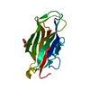
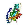
| Title | Outer Surface Protein B of B. burgdorferi: crystal structure of the C-terminal fragment | ||||||
 Components Components | Outer surface protein B | ||||||
 Keywords Keywords | MEMBRANE PROTEIN / INTERMOLECULAR BETA SHEET / EXTENDED BETA SHEET | ||||||
| Function / homology |  Function and homology information Function and homology information | ||||||
| Biological species |  Borrelia burgdorferi (Lyme disease spirochete) Borrelia burgdorferi (Lyme disease spirochete) | ||||||
| Method |  X-RAY DIFFRACTION / SYNCHROTRON / MOLECULAR REPLACEMENT / Resolution: 2 Å X-RAY DIFFRACTION / SYNCHROTRON / MOLECULAR REPLACEMENT / Resolution: 2 Å | ||||||
 Authors Authors | Becker, M. / Bunikis, J. / Lade, B.D. / Dunn, J.J. / Barbour, A.G. / Lawson, C.L. | ||||||
 Citation Citation | Journal: J.Biol.Chem. / Year: 2005 Title: Structural Investigation of Borrelia burgdorferi OspB, a BactericidalFab Target. Authors: Becker, M. / Bunikis, J. / Lade, B.D. / Dunn, J.J. / Barbour, A.G. / Lawson, C.L. | ||||||
| History |
| ||||||
| Remark 650 | HELIX DETERMINATION METHOD: AUTHOR |
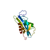
- Structure visualization
| Structure viewer | Molecule: MolmilJmol/JSmol |
|---|
- Downloads & links
-Download
| PDBx/mmCIF format | 1p4p.cif.gz | 41.2 KB | Display | PDBx/mmCIF format |
|---|---|---|---|---|
| PDB format | pdb1p4p.ent.gz | 28.6 KB | Display | PDB format |
| PDBx/mmJSON format | 1p4p.json.gz | Tree view | PDBx/mmJSON format | |
| Others |  Other downloads Other downloads |
-Validation report
| Arichive directory | https://data.pdbj.org/pub/pdb/validation_reports/p4/1p4pftp://data.pdbj.org/pub/pdb/validation_reports/p4/1p4p | HTTPS FTP |
|---|
-Related structure data
| Related structure data | 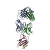 1rjlC 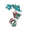 1ospS S: Starting model for refinement C: citing same article ( |
|---|---|
| Similar structure data |


-Links
 PDBj
PDBj


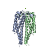
- Assembly
| Deposited unit | 
| ||||||||
|---|---|---|---|---|---|---|---|---|---|
| 1 |
| ||||||||
| Unit cell |
|
-Components
| #1: Protein | Mass: 15653.579 Da / Num. of mol.: 1 / Fragment: OspB C-terminal fragment Source method: isolated from a genetically manipulated source Source: (gene. exp.) Borrelia burgdorferi (Lyme disease spirochete)Gene: OSPB / Plasmid: pET9c / Production host: |
|---|---|
| #2: Water | ChemComp-HOH /  Mass: 18.015 Da / Num. of mol.: 118 / Source method: isolated from a natural source / Formula: H2O Mass: 18.015 Da / Num. of mol.: 118 / Source method: isolated from a natural source / Formula: H2O |
-Experimental details
-Experiment
| Experiment | Method: X-RAY DIFFRACTION / Number of used crystals: 1 |
|---|
- Sample preparation
| Crystal | Density Matthews: 2.02 Å3/Da / Density % sol: 39.05 % |
|---|---|
| Crystal grow | Temperature: 295 K / Method: vapor diffusion, hanging drop / pH: 9.5 Details: PEG550MME, Bicine, sodium bromide, sodium citrate, pH 9.5, VAPOR DIFFUSION, HANGING DROP, temperature 295K |
-Data collection
| Diffraction | Mean temperature: 283 K | |||||||||
|---|---|---|---|---|---|---|---|---|---|---|
| Diffraction source | Source: SYNCHROTRON / Site: NSLS  / Beamline: X12B / Wavelength: 0.979, 1.073 / Beamline: X12B / Wavelength: 0.979, 1.073 | |||||||||
| Detector | Type: MARRESEARCH / Detector: IMAGE PLATE / Date: Mar 1, 1997 / Details: focusing mirror | |||||||||
| Radiation | Monochromator: two-crystal Si (111) / Protocol: SINGLE WAVELENGTH / Monochromatic (M) / Laue (L): M / Scattering type: x-ray | |||||||||
| Radiation wavelength |
| |||||||||
| Reflection | Resolution: 2→50 Å / Num. all: 9012 / Num. obs: 8090 / % possible obs: 89.8 % / Observed criterion σ(F): -10 / Observed criterion σ(I): -10 / Redundancy: 23.7 % / Rmerge(I) obs: 0.063 / Net I/σ(I): 34.1 | |||||||||
| Reflection shell | Resolution: 2→2.03 Å / Rmerge(I) obs: 0.206 / Mean I/σ(I) obs: 12.6 / % possible all: 77.9 |
- Processing
| Software |
| |||||||||||||||||||||||||
|---|---|---|---|---|---|---|---|---|---|---|---|---|---|---|---|---|---|---|---|---|---|---|---|---|---|---|
| Refinement | Method to determine structure: MOLECULAR REPLACEMENT Starting model: PDB ENTRY 1OSP Resolution: 2→30 Å / Isotropic thermal model: anisotropic / Cross valid method: THROUGHOUT / σ(F): 0 / Stereochemistry target values: Engh & Huber Details: Used CNS for simulated annealing, followed by conjugate gradient minimization against the maximum likelihood function. Then ran Refmac, and followed with 1 step of B-factor refinement in CNS. ...Details: Used CNS for simulated annealing, followed by conjugate gradient minimization against the maximum likelihood function. Then ran Refmac, and followed with 1 step of B-factor refinement in CNS. The loop from residue 218 to residue 221 (LYS ASP GLY LYS) is poorly ordered (is not clearly defined in the density).
| |||||||||||||||||||||||||
| Displacement parameters | Biso mean: 32.3 Å2
| |||||||||||||||||||||||||
| Refine analyze | Luzzati coordinate error obs: 0.247 Å | |||||||||||||||||||||||||
| Refinement step | Cycle: LAST / Resolution: 2→30 Å
| |||||||||||||||||||||||||
| Refine LS restraints |
|
