 Movie
Movie Controller

[English] 日本語
 Yorodumi
- PDB-1quu: CRYSTAL STRUCTURE OF TWO CENTRAL SPECTRIN-LIKE REPEATS FROM ALPHA... -
+ Open data

- Basic information
| Entry | Database: PDB / ID: 1quu | ||||||
|---|---|---|---|---|---|---|---|
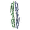
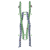
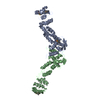
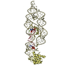
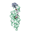
| Title | CRYSTAL STRUCTURE OF TWO CENTRAL SPECTRIN-LIKE REPEATS FROM ALPHA-ACTININ | ||||||
 Components Components | HUMAN SKELETAL MUSCLE ALPHA-ACTININ 2 | ||||||
 Keywords Keywords | CONTRACTILE PROTEIN / TRIPLE-HELIX COILED COIL | ||||||
| Function / homology |  Function and homology information Function and homology informationactin filament uncapping / FATZ binding / titin Z domain binding / positive regulation of endocytic recycling / phospholipase C-activating angiotensin-activated signaling pathway / positive regulation of cation channel activity / negative regulation of protein localization to cell surface / channel activator activity / LIM domain binding / microspike assembly ...actin filament uncapping / FATZ binding / titin Z domain binding / positive regulation of endocytic recycling / phospholipase C-activating angiotensin-activated signaling pathway / positive regulation of cation channel activity / negative regulation of protein localization to cell surface / channel activator activity / LIM domain binding / microspike assembly / positive regulation of potassium ion transport / focal adhesion assembly / structural constituent of postsynaptic actin cytoskeleton / muscle cell development / postsynaptic actin cytoskeleton / Striated Muscle Contraction / Nephrin family interactions / Assembly and cell surface presentation of NMDA receptors / cardiac muscle cell development / sarcomere organization / structural constituent of muscle / cortical actin cytoskeleton / pseudopodium / Negative regulation of NMDA receptor-mediated neuronal transmission / Unblocking of NMDA receptors, glutamate binding and activation / negative regulation of potassium ion transport / Long-term potentiation / postsynaptic density, intracellular component / cytoskeletal protein binding / titin binding / phosphatidylinositol-4,5-bisphosphate binding / Ras activation upon Ca2+ influx through NMDA receptor / platelet alpha granule lumen / cell projection / protein localization to plasma membrane / actin filament / filopodium / regulation of membrane potential / postsynaptic density membrane / integrin binding / Z disc / actin filament binding / cell junction / Platelet degranulation / RAF/MAP kinase cascade / actin cytoskeleton organization / regulation of apoptotic process / dendritic spine / transmembrane transporter binding / cytoskeleton / transcription coactivator activity / cell adhesion / protein domain specific binding / focal adhesion / calcium ion binding / glutamatergic synapse / extracellular exosome / extracellular region / identical protein binding / cytosol Similarity search - Function | ||||||
| Biological species |  Homo sapiens (human) Homo sapiens (human) | ||||||
| Method |  X-RAY DIFFRACTION / SYNCHROTRON / Resolution: 2.5 Å X-RAY DIFFRACTION / SYNCHROTRON / Resolution: 2.5 Å | ||||||
 Authors Authors | Djinovic-Carugo, K. / Young, P. / Gautel, M. / Saraste, M. | ||||||
 Citation Citation | Journal: Cell(Cambridge,Mass.) / Year: 1999 Title: Structure of the alpha-actinin rod: molecular basis for cross-linking of actin filaments. Authors: Djinovic-Carugo, K. / Young, P. / Gautel, M. / Saraste, M. | ||||||
| History |
|
- Structure visualization
| Structure viewer | Molecule: MolmilJmol/JSmol |
|---|
- Downloads & links
-Download
| PDBx/mmCIF format | 1quu.cif.gz | 66.1 KB | Display | PDBx/mmCIF format |
|---|---|---|---|---|
| PDB format | pdb1quu.ent.gz | 49.7 KB | Display | PDB format |
| PDBx/mmJSON format | 1quu.json.gz | Tree view | PDBx/mmJSON format | |
| Others |  Other downloads Other downloads |
-Validation report
| Arichive directory | https://data.pdbj.org/pub/pdb/validation_reports/qu/1quuftp://data.pdbj.org/pub/pdb/validation_reports/qu/1quu | HTTPS FTP |
|---|
-Related structure data
| Similar structure data |
|---|


-Links
 PDBj
PDBj


- Assembly
| Deposited unit | 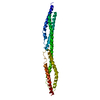
| ||||||||
|---|---|---|---|---|---|---|---|---|---|
| 1 | 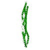
| ||||||||
| Unit cell |
|
-Components
| #1: Protein | Mass: 29283.828 Da / Num. of mol.: 1 Fragment: SPECTRIN-LIKE REPEATS 2 AND 3 - AMINO ACIDS 391 TO 637 Source method: isolated from a genetically manipulated source Source: (gene. exp.) Homo sapiens (human) / Tissue: SKELETAL MUSCLE / Plasmid: PET 8C / Production host:  |
|---|---|
| #2: Water | ChemComp-HOH /  Mass: 18.015 Da / Num. of mol.: 151 / Source method: isolated from a natural source / Formula: H2O Mass: 18.015 Da / Num. of mol.: 151 / Source method: isolated from a natural source / Formula: H2O |
-Experimental details
-Experiment
| Experiment | Method: X-RAY DIFFRACTION / Number of used crystals: 1 |
|---|
- Sample preparation
| Crystal | Density Matthews: 3.47 Å3/Da / Density % sol: 64.58 % | |||||||||||||||||||||||||||||||||||||||||||||||||
|---|---|---|---|---|---|---|---|---|---|---|---|---|---|---|---|---|---|---|---|---|---|---|---|---|---|---|---|---|---|---|---|---|---|---|---|---|---|---|---|---|---|---|---|---|---|---|---|---|---|---|
| Crystal grow | Temperature: 277 K / Method: vapor diffusion, hanging drop / pH: 7.5 Details: 26% PEG 400 (FLUKA), 100 MM MGSO4, 100 MM HEPES OR TRIS-HCL, pH 7.5, VAPOR DIFFUSION, HANGING DROP | |||||||||||||||||||||||||||||||||||||||||||||||||
| Crystal grow | *PLUS pH: 7.4 / Method: vapor diffusion | |||||||||||||||||||||||||||||||||||||||||||||||||
| Components of the solutions | *PLUS
|
-Data collection
| Diffraction | Mean temperature: 100 K |
|---|---|
| Diffraction source | Source: SYNCHROTRON / Site: ESRF  / Beamline: ID14-3 / Wavelength: 0.947 / Beamline: ID14-3 / Wavelength: 0.947 |
| Detector | Type: OFF-LINE SCANNER / Detector: IMAGE PLATE / Date: Feb 10, 1998 |
| Radiation | Protocol: SINGLE WAVELENGTH / Monochromatic (M) / Laue (L): M / Scattering type: x-ray |
| Radiation wavelength | Wavelength: 0.947 Å / Relative weight: 1 |
| Reflection | Resolution: 2.2→25 Å / Num. all: 21275 / Num. obs: 21275 / % possible obs: 94.6 % / Observed criterion σ(F): 0 / Observed criterion σ(I): 0 / Redundancy: 6.1 % / Biso Wilson estimate: 48.6 Å2 / Rmerge(I) obs: 0.062 / Net I/σ(I): 15.3 |
| Reflection shell | Resolution: 2.2→2.24 Å / Redundancy: 2.8 % / Rmerge(I) obs: 0.339 / % possible all: 89.8 |
| Reflection | *PLUS |
| Reflection shell | *PLUS % possible obs: 89.8 % |
- Processing
| Software |
| ||||||||||||||||||||||||||||||||||||||||||||||||||||||||||||
|---|---|---|---|---|---|---|---|---|---|---|---|---|---|---|---|---|---|---|---|---|---|---|---|---|---|---|---|---|---|---|---|---|---|---|---|---|---|---|---|---|---|---|---|---|---|---|---|---|---|---|---|---|---|---|---|---|---|---|---|---|---|
| Refinement | Resolution: 2.5→25 Å / σ(F): 2 / σ(I): 0 / Stereochemistry target values: ENGH & HUBER
| ||||||||||||||||||||||||||||||||||||||||||||||||||||||||||||
| Refinement step | Cycle: LAST / Resolution: 2.5→25 Å
| ||||||||||||||||||||||||||||||||||||||||||||||||||||||||||||
| Refine LS restraints |
| ||||||||||||||||||||||||||||||||||||||||||||||||||||||||||||
| Software | *PLUS Name: CNS / Classification: refinement | ||||||||||||||||||||||||||||||||||||||||||||||||||||||||||||
| Refinement | *PLUS Highest resolution: 2.5 Å / Lowest resolution: 25 Å / σ(F): 2 | ||||||||||||||||||||||||||||||||||||||||||||||||||||||||||||
| Solvent computation | *PLUS | ||||||||||||||||||||||||||||||||||||||||||||||||||||||||||||
| Displacement parameters | *PLUS Biso mean: 57.2 Å2 |
