 ムービー
ムービー コントローラー


+ データを開く

- 基本情報
基本情報
| 登録情報 | データベース: PDB / ID: 1quu | ||||||
|---|---|---|---|---|---|---|---|
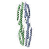
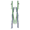
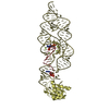
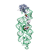
| タイトル | CRYSTAL STRUCTURE OF TWO CENTRAL SPECTRIN-LIKE REPEATS FROM ALPHA-ACTININ | ||||||
 要素 要素 | HUMAN SKELETAL MUSCLE ALPHA-ACTININ 2 | ||||||
 キーワード キーワード |  CONTRACTILE PROTEIN / TRIPLE-HELIX COILED COIL CONTRACTILE PROTEIN / TRIPLE-HELIX COILED COIL | ||||||
| 機能・相同性 |  機能・相同性情報 機能・相同性情報actin filament uncapping / FATZ binding / titin Z domain binding / phospholipase C-activating angiotensin-activated signaling pathway / positive regulation of endocytic recycling / positive regulation of potassium ion transmembrane transporter activity / negative regulation of potassium ion transmembrane transporter activity / positive regulation of cation channel activity / LIM domain binding / negative regulation of protein localization to cell surface ...actin filament uncapping / FATZ binding / titin Z domain binding / phospholipase C-activating angiotensin-activated signaling pathway / positive regulation of endocytic recycling / positive regulation of potassium ion transmembrane transporter activity / negative regulation of potassium ion transmembrane transporter activity / positive regulation of cation channel activity / LIM domain binding / negative regulation of protein localization to cell surface / microspike assembly / postsynaptic actin cytoskeleton / muscle cell development / positive regulation of potassium ion transport / focal adhesion assembly / Striated Muscle Contraction / Assembly and cell surface presentation of NMDA receptors / cardiac muscle cell development / Nephrin family interactions / structural constituent of muscle / sarcomere organization / cortical actin cytoskeleton / Negative regulation of NMDA receptor-mediated neuronal transmission / Unblocking of NMDA receptors, glutamate binding and activation / 仮足 / postsynaptic density, intracellular component / negative regulation of potassium ion transport / 長期増強 / titin binding / phosphatidylinositol-4,5-bisphosphate binding / Ras activation upon Ca2+ influx through NMDA receptor / regulation of membrane potential / cytoskeletal protein binding / nuclear receptor coactivator activity / platelet alpha granule lumen / filopodium / cell projection / マイクロフィラメント / protein localization to plasma membrane / postsynaptic density membrane / Z disc / actin filament binding / integrin binding / Platelet degranulation / 細胞結合 / actin cytoskeleton organization / RAF/MAP kinase cascade / regulation of apoptotic process / transmembrane transporter binding / 樹状突起スパイン / 細胞骨格 / 細胞接着 / protein domain specific binding / focal adhesion / glutamatergic synapse / calcium ion binding / extracellular exosome / extracellular region / identical protein binding / 細胞質基質類似検索 - 分子機能 | ||||||
| 生物種 |  Homo sapiens (ヒト) Homo sapiens (ヒト) | ||||||
| 手法 | X線回折 / シンクロトロン / 解像度: 2.5 Å | ||||||
 データ登録者 データ登録者 | Djinovic-Carugo, K. / Young, P. / Gautel, M. / Saraste, M. | ||||||
 引用 引用 | ジャーナル: Cell(Cambridge,Mass.) / 年: 1999 タイトル: Structure of the alpha-actinin rod: molecular basis for cross-linking of actin filaments. 著者: Djinovic-Carugo, K. / Young, P. / Gautel, M. / Saraste, M. | ||||||
| 履歴 |
|
- 構造の表示
構造の表示
| 構造ビューア | 分子: MolmilJmol/JSmol |
|---|
- ダウンロードとリンク
-ダウンロード
| PDBx/mmCIF形式 | 1quu.cif.gz | 66.1 KB | 表示 | PDBx/mmCIF形式 |
|---|---|---|---|---|
| PDB形式 | pdb1quu.ent.gz | 49.7 KB | 表示 | PDB形式 |
| PDBx/mmJSON形式 | 1quu.json.gz | ツリー表示 | PDBx/mmJSON形式 | |
| その他 |  その他のダウンロード その他のダウンロード |
-検証レポート
| アーカイブディレクトリ | https://data.pdbj.org/pub/pdb/validation_reports/qu/1quuftp://data.pdbj.org/pub/pdb/validation_reports/qu/1quu | HTTPS FTP |
|---|
-関連構造データ

-リンク
 PDBj
PDBj


- 集合体
集合体
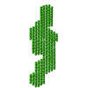
| 登録構造単位 | 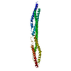
| ||||||||
|---|---|---|---|---|---|---|---|---|---|
| 1 | 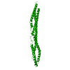
| ||||||||
| 単位格子 |
|
-要素
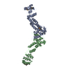
| #1: タンパク質 | 分子量: 29283.828 Da / 分子数: 1 断片: SPECTRIN-LIKE REPEATS 2 AND 3 - AMINO ACIDS 391 TO 637 由来タイプ: 組換発現 / 由来: (組換発現) Homo sapiens (ヒト) / 組織: SKELETAL MUSCLE骨格筋 / プラスミド: PET 8C / 発現宿主:  Escherichia coli (大腸菌) / 参照: UniProt: P35609 Escherichia coli (大腸菌) / 参照: UniProt: P35609 |
|---|---|
| #2: 水 | ChemComp-HOH / 水 分子量: 18.015 Da / 分子数: 151 / 由来タイプ: 天然 / 式: H2O 分子量: 18.015 Da / 分子数: 151 / 由来タイプ: 天然 / 式: H2O |
-実験情報
-実験
| 実験 | 手法: X線回折 / 使用した結晶の数: 1 |
|---|
- 試料調製
試料調製
| 結晶 | マシュー密度: 3.47 Å3/Da / 溶媒含有率: 64.58 % | |||||||||||||||||||||||||||||||||||||||||||||||||
|---|---|---|---|---|---|---|---|---|---|---|---|---|---|---|---|---|---|---|---|---|---|---|---|---|---|---|---|---|---|---|---|---|---|---|---|---|---|---|---|---|---|---|---|---|---|---|---|---|---|---|
| 結晶化 | 温度: 277 K / 手法: 蒸気拡散法, ハンギングドロップ法 / pH: 7.5 詳細: 26% PEG 400 (FLUKA), 100 MM MGSO4, 100 MM HEPES OR TRIS-HCL, pH 7.5, VAPOR DIFFUSION, HANGING DROP | |||||||||||||||||||||||||||||||||||||||||||||||||
| 結晶化 | *PLUS pH: 7.4 / 手法: 蒸気拡散法 | |||||||||||||||||||||||||||||||||||||||||||||||||
| 溶液の組成 | *PLUS
|
-データ収集
| 回折 | 平均測定温度: 100 K |
|---|---|
| 放射光源 | 由来: シンクロトロン / サイト: ESRF  / ビームライン: ID14-3 / 波長: 0.947 / ビームライン: ID14-3 / 波長: 0.947 |
| 検出器 | タイプ: OFF-LINE SCANNER / 検出器: IMAGE PLATE / 日付: 1998年2月10日 |
| 放射 | プロトコル: SINGLE WAVELENGTH / 単色(M)・ラウエ(L): M / 散乱光タイプ: x-ray |
| 放射波長 | 波長: 0.947 Å / 相対比: 1 |
| 反射 | 解像度: 2.2→25 Å / Num. all: 21275 / Num. obs: 21275 / % possible obs: 94.6 % / Observed criterion σ(F): 0 / Observed criterion σ(I): 0 / 冗長度: 6.1 % / Biso Wilson estimate: 48.6 Å2 / Rmerge(I) obs: 0.062 / Net I/σ(I): 15.3 |
| 反射 シェル | 解像度: 2.2→2.24 Å / 冗長度: 2.8 % / Rmerge(I) obs: 0.339 / % possible all: 89.8 |
| 反射 | *PLUS |
| 反射 シェル | *PLUS % possible obs: 89.8 % |
- 解析
解析
| ソフトウェア |
| ||||||||||||||||||||||||||||||||||||||||||||||||||||||||||||
|---|---|---|---|---|---|---|---|---|---|---|---|---|---|---|---|---|---|---|---|---|---|---|---|---|---|---|---|---|---|---|---|---|---|---|---|---|---|---|---|---|---|---|---|---|---|---|---|---|---|---|---|---|---|---|---|---|---|---|---|---|---|
| 精密化 | 解像度: 2.5→25 Å / σ(F): 2 / σ(I): 0 / 立体化学のターゲット値: ENGH & HUBER
| ||||||||||||||||||||||||||||||||||||||||||||||||||||||||||||
| 精密化ステップ | サイクル: LAST / 解像度: 2.5→25 Å
| ||||||||||||||||||||||||||||||||||||||||||||||||||||||||||||
| 拘束条件 |
| ||||||||||||||||||||||||||||||||||||||||||||||||||||||||||||
| ソフトウェア | *PLUS 名称: CNS / 分類: refinement | ||||||||||||||||||||||||||||||||||||||||||||||||||||||||||||
| 精密化 | *PLUS 最高解像度: 2.5 Å / 最低解像度: 25 Å / σ(F): 2 | ||||||||||||||||||||||||||||||||||||||||||||||||||||||||||||
| 溶媒の処理 | *PLUS | ||||||||||||||||||||||||||||||||||||||||||||||||||||||||||||
| 原子変位パラメータ | *PLUS Biso mean: 57.2 Å2 |
