 Movie
Movie Controller

[English] 日本語
 Yorodumi
- PDB-1qkf: SOLUTION STRUCTURE OF THE RIBOSOMAL PROTEIN S19 FROM THERMUS THER... -
+ Open data

- Basic information
| Entry | Database: PDB / ID: 1qkf | ||||||
|---|---|---|---|---|---|---|---|
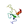
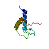
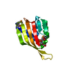
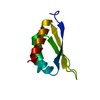
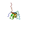
| Title | SOLUTION STRUCTURE OF THE RIBOSOMAL PROTEIN S19 FROM THERMUS THERMOPHILUS | ||||||
 Components Components | 30S RIBOSOMAL PROTEIN S19 | ||||||
 Keywords Keywords | RIBOSOMAL PROTEIN / RIBOSOME / THERMUS THERMOPHILUS / S19 | ||||||
| Function / homology |  Function and homology information Function and homology informationribosomal small subunit assembly / cytosolic small ribosomal subunit / rRNA binding / structural constituent of ribosome / translation Similarity search - Function | ||||||
| Biological species |   THERMUS THERMOPHILUS (bacteria) THERMUS THERMOPHILUS (bacteria) | ||||||
| Method | SOLUTION NMR / simulated annealing | ||||||
| Model type details | MINIMIZED AVERAGE | ||||||
 Authors Authors | Helgstrand, M. / Rak, A.V. / Allard, P. / Davydova, N. / Garber, M.B. / Hard, T. | ||||||
 Citation Citation | Journal: J. Mol. Biol. / Year: 1999 Title: Solution structure of the ribosomal protein S19 from Thermus thermophilus. Authors: Helgstrand, M. / Rak, A.V. / Allard, P. / Davydova, N. / Garber, M.B. / Hard, T. | ||||||
| History |
| ||||||
| Remark 650 | HELIX DETERMINATION METHOD: KABSCH AND SANDER | ||||||
| Remark 700 | SHEET DETERMINATION METHOD: KABSCH AND SANDER |
- Structure visualization
| Structure viewer | Molecule: MolmilJmol/JSmol |
|---|
- Downloads & links
-Download
| PDBx/mmCIF format | 1qkf.cif.gz | 37.4 KB | Display | PDBx/mmCIF format |
|---|---|---|---|---|
| PDB format | pdb1qkf.ent.gz | 26.1 KB | Display | PDB format |
| PDBx/mmJSON format | 1qkf.json.gz | Tree view | PDBx/mmJSON format | |
| Others |  Other downloads Other downloads |
-Validation report
| Summary document | 1qkf_validation.pdf.gz | 336.5 KB | Display | wwPDB validaton report |
|---|---|---|---|---|
| Full document | 1qkf_full_validation.pdf.gz | 343 KB | Display | |
| Data in XML | 1qkf_validation.xml.gz | 3.8 KB | Display | |
| Data in CIF | 1qkf_validation.cif.gz | 4.8 KB | Display | |
| Arichive directory | https://data.pdbj.org/pub/pdb/validation_reports/qk/1qkfftp://data.pdbj.org/pub/pdb/validation_reports/qk/1qkf | HTTPS FTP |
-Related structure data


-Links
 PDBj
PDBj


- Assembly
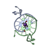
| Deposited unit | 
| |||||||||
|---|---|---|---|---|---|---|---|---|---|---|
| 1 |
| |||||||||
| NMR ensembles |
|
-Components
| #1: Protein | Mass: 10474.269 Da / Num. of mol.: 1 Source method: isolated from a genetically manipulated source Source: (gene. exp.) THERMUS THERMOPHILUS (bacteria) / Plasmid: PTTHS19 / Production host: |
|---|
-Experimental details
-Experiment
| Experiment | Method: SOLUTION NMR | ||||||||||||||||||||||||||||||||||||||||||||||||||||||||
|---|---|---|---|---|---|---|---|---|---|---|---|---|---|---|---|---|---|---|---|---|---|---|---|---|---|---|---|---|---|---|---|---|---|---|---|---|---|---|---|---|---|---|---|---|---|---|---|---|---|---|---|---|---|---|---|---|---|
| NMR experiment |
| ||||||||||||||||||||||||||||||||||||||||||||||||||||||||
| NMR details | Text: MINIMIZED AVERAGE STRUCTURE. |
 HNCA
HNCA- Sample preparation
| Details | Contents: 90% WATER/10% D2O |
|---|---|
| Sample conditions | Ionic strength: 0.250 M / pH: 6.5 / Pressure: 1 atm / Temperature: 303 K |
| Crystal grow | *PLUS Method: other / Details: NMR |
-NMR measurement
| NMR spectrometer |
|
|---|
- Processing
| NMR software |
| ||||||||||||||||||||||||||||
|---|---|---|---|---|---|---|---|---|---|---|---|---|---|---|---|---|---|---|---|---|---|---|---|---|---|---|---|---|---|
| Refinement | Method: simulated annealing / Software ordinal: 1 Details: THE STRUCTURE WAS DETERMINED USING 1104 DISTANCE RESTRAINTS, 42 DIHEDRAL ANGLE RESTRAINTS AND 14 HYDROGEN BOND RESTRAINTS. 50 STRUCTURES WERE CALCULATED AND REFINED USING AN AB INITIO ...Details: THE STRUCTURE WAS DETERMINED USING 1104 DISTANCE RESTRAINTS, 42 DIHEDRAL ANGLE RESTRAINTS AND 14 HYDROGEN BOND RESTRAINTS. 50 STRUCTURES WERE CALCULATED AND REFINED USING AN AB INITIO SIMULATED ANNEALING PROTOCOL FOR X- PLOR AND THEN REFINED IN TWO STEPS. AN R-6 AVERAGING PROTOCOL WAS USED FOR NON-STEREOSPECIFICALLY ASSIGNED PROTONS [1]. DURING THE SIMULATED ANNEALING STEP AND THE FIRST REFINEMENT STEP ONLY THE REPULSIVE PART OF THE VAN DER WAALS INTERACTION WAS INCLUDED. IN THE SECOND REFINEMENT STEP THE VAN DER WAALS INTERACTION WAS PARAMETERIZED USING A LENNARD-JONES POTENTIAL INCLUDING THE ATTRACTIVE PART. 21 STRUCTURES WERE SELECTED ON THE BASIS OF CUMULATIVE RMSD VALUES OF STRUCTURES, ORDERED AFTER OVERALL ENERGY, AND RAMACHANDRAN BEHAVIOR FOR REGIONS WITH LOW RESTRAINT DENSITIES. [1] BRUNGER, A. T., CLORE, G. M., GRONENBORN, A. M. & KARPLUS, M. (1986). THREE-DIMENSIONAL STRUCTURE OF PROTEINS DETERMINED BY MOLECULAR DYNAMICS WITH INTERPROTON DISTANCE RESTRAINTS: APPLICATION TO CRAMBIN. PROC NATL ACAD SCI USA 83, 3801-3805. OTHER DETAILS OF STRUCTURE REFINEMENT CAN BE FOUND IN THE JRNL CITATION. | ||||||||||||||||||||||||||||
| NMR ensemble | Conformer selection criteria: CUMULATIVE RMSD OF STRUCTURES SORTED AFTER TOTAL ENERGY Conformers calculated total number: 21 / Conformers submitted total number: 1 |
