 Movie
Movie Controller

[English] 日本語
 Yorodumi
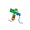
- PDB-3nx6: Crystal Structure of co-chaperonin, GroES (Xoo4289) from Xanthomo... -
+ Open data

- Basic information
| Entry | Database: PDB / ID: 3nx6 | ||||||
|---|---|---|---|---|---|---|---|
| Title | Crystal Structure of co-chaperonin, GroES (Xoo4289) from Xanthomonas oryzae pv. oryzae KACC10331 | ||||||
 Components Components | 10kDa chaperonin | ||||||
 Keywords Keywords | CHAPERONE / Bacterial blight / Xoo4289 / GroES / Xanthomonas oryzae pv. oryzae KACC10331 | ||||||
| Function / homology |  Function and homology information Function and homology informationprotein folding chaperone / : / protein-folding chaperone binding / ATP binding / metal ion binding / cytoplasm Similarity search - Function | ||||||
| Biological species |  Xanthomonas oryzae pv. oryzae (bacteria) Xanthomonas oryzae pv. oryzae (bacteria) | ||||||
| Method |  X-RAY DIFFRACTION / SYNCHROTRON / MOLECULAR REPLACEMENT / Resolution: 1.97 Å X-RAY DIFFRACTION / SYNCHROTRON / MOLECULAR REPLACEMENT / Resolution: 1.97 Å | ||||||
 Authors Authors | Natarajan, S. / Doan, T.T.N. / Kang, L.-W. | ||||||
 Citation Citation | Journal: to be published Title: Crystal Structure of co-chaperonin, GroES (Xoo4289) from Xanthomonas oryzae pv. oryzae KACC10331 Authors: Natarajan, S. / Doan, T.T.N. / Kang, L.-W. | ||||||
| History |
|
- Structure visualization
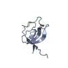
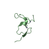
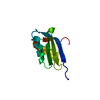
| Structure viewer | Molecule: MolmilJmol/JSmol |
|---|
- Downloads & links
-Download
| PDBx/mmCIF format | 3nx6.cif.gz | 26.6 KB | Display | PDBx/mmCIF format |
|---|---|---|---|---|
| PDB format | pdb3nx6.ent.gz | 17.5 KB | Display | PDB format |
| PDBx/mmJSON format | 3nx6.json.gz | Tree view | PDBx/mmJSON format | |
| Others |  Other downloads Other downloads |
-Validation report
| Arichive directory | https://data.pdbj.org/pub/pdb/validation_reports/nx/3nx6ftp://data.pdbj.org/pub/pdb/validation_reports/nx/3nx6 | HTTPS FTP |
|---|
-Related structure data
| Related structure data | 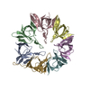 1wnrS S: Starting model for refinement |
|---|---|
| Similar structure data |


-Links
 PDBj
PDBj

- Assembly
| Deposited unit | 
| ||||||||
|---|---|---|---|---|---|---|---|---|---|
| 1 |
| ||||||||
| Unit cell |
| ||||||||
| Details | IN SOLUTION IT COULD FORM HEPTAMER. |
-Components
| #1: Protein | Mass: 9974.401 Da / Num. of mol.: 1 / Fragment: UNP residues 40-134 / Mutation: R55H Source method: isolated from a genetically manipulated source Source: (gene. exp.) Xanthomonas oryzae pv. oryzae (bacteria)Strain: KACC10331 / Gene: Xoo4289 / Plasmid: pET11a / Production host: |
|---|---|
| #2: Water | ChemComp-HOH /  Mass: 18.015 Da / Num. of mol.: 37 / Source method: isolated from a natural source / Formula: H2O Mass: 18.015 Da / Num. of mol.: 37 / Source method: isolated from a natural source / Formula: H2O |
-Experimental details
-Experiment
| Experiment | Method: X-RAY DIFFRACTION / Number of used crystals: 1 |
|---|
- Sample preparation
| Crystal | Density Matthews: 2.19 Å3/Da / Density % sol: 43.87 % |
|---|---|
| Crystal grow | Temperature: 287 K / Method: vapor diffusion, hanging drop / pH: 4.1 Details: 0.2M Sodium citrate pH 4.1, 16% PEG 400, 0.2M Li2SO4, VAPOR DIFFUSION, HANGING DROP, temperature 287K |
-Data collection
| Diffraction | Mean temperature: 100 K |
|---|---|
| Diffraction source | Source: SYNCHROTRON / Site: PAL/PLS  / Beamline: 4A / Wavelength: 1 Å / Beamline: 4A / Wavelength: 1 Å |
| Detector | Type: ADSC QUANTUM 210 / Detector: CCD / Date: Jun 11, 2010 |
| Radiation | Protocol: SINGLE WAVELENGTH / Monochromatic (M) / Laue (L): M / Scattering type: x-ray |
| Radiation wavelength | Wavelength: 1 Å / Relative weight: 1 |
| Reflection | Resolution: 1.97→55.78 Å / Num. all: 6225 / Num. obs: 5590 / % possible obs: 94.52 % / Observed criterion σ(F): 1 / Observed criterion σ(I): 1 |
| Reflection shell | Resolution: 1.97→2.08 Å / % possible all: 98.9 |
- Processing
| Software |
| |||||||||||||||||||||||||||||||||||||||||||||||||||||||||||||||||||||||||||||||||||||
|---|---|---|---|---|---|---|---|---|---|---|---|---|---|---|---|---|---|---|---|---|---|---|---|---|---|---|---|---|---|---|---|---|---|---|---|---|---|---|---|---|---|---|---|---|---|---|---|---|---|---|---|---|---|---|---|---|---|---|---|---|---|---|---|---|---|---|---|---|---|---|---|---|---|---|---|---|---|---|---|---|---|---|---|---|---|---|
| Refinement | Method to determine structure: MOLECULAR REPLACEMENT Starting model: PDB ENTRY 1WNR Resolution: 1.97→32.21 Å / Cor.coef. Fo:Fc: 0.957 / Cor.coef. Fo:Fc free: 0.912 / SU B: 6.215 / SU ML: 0.163 / Cross valid method: THROUGHOUT / ESU R Free: 0.195 / Stereochemistry target values: MAXIMUM LIKELIHOOD / Details: HYDROGENS HAVE BEEN ADDED IN THE RIDING POSITIONS
| |||||||||||||||||||||||||||||||||||||||||||||||||||||||||||||||||||||||||||||||||||||
| Solvent computation | Ion probe radii: 0.8 Å / Shrinkage radii: 0.8 Å / VDW probe radii: 1.2 Å / Solvent model: MASK | |||||||||||||||||||||||||||||||||||||||||||||||||||||||||||||||||||||||||||||||||||||
| Displacement parameters | Biso mean: 36.821 Å2
| |||||||||||||||||||||||||||||||||||||||||||||||||||||||||||||||||||||||||||||||||||||
| Refinement step | Cycle: LAST / Resolution: 1.97→32.21 Å
| |||||||||||||||||||||||||||||||||||||||||||||||||||||||||||||||||||||||||||||||||||||
| Refine LS restraints |
| |||||||||||||||||||||||||||||||||||||||||||||||||||||||||||||||||||||||||||||||||||||
| LS refinement shell | Resolution: 1.974→2.025 Å / Total num. of bins used: 20
|
