 Movie
Movie Controller


+ Open data

- Basic information
| Entry | Database: PDB / ID: 1wnr | ||||||
|---|---|---|---|---|---|---|---|
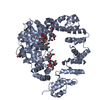
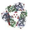
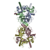
| Title | Crystal structure of the Cpn10 from Thermus thermophilus HB8 | ||||||
 Components Components | 10 kDa chaperonin | ||||||
 Keywords Keywords | CHAPERONE / CO-Chaperonin / protein CPN10 / Groes / Thermus Thermophilus / RIKEN Structural Genomics/Proteomics Initiative / RSGI / Structural Genomics | ||||||
| Function / homology |  Function and homology information Function and homology informationprotein folding chaperone / : / protein-folding chaperone binding / ATP binding / metal ion binding / cytoplasm Similarity search - Function | ||||||
| Biological species |   Thermus thermophilus (bacteria) Thermus thermophilus (bacteria) | ||||||
| Method |  X-RAY DIFFRACTION / SYNCHROTRON / MOLECULAR REPLACEMENT / Resolution: 2.9 Å X-RAY DIFFRACTION / SYNCHROTRON / MOLECULAR REPLACEMENT / Resolution: 2.9 Å | ||||||
 Authors Authors | Numoto, N. / Kita, A. / Miki, K. / RIKEN Structural Genomics/Proteomics Initiative (RSGI) | ||||||
 Citation Citation | Journal: Proteins / Year: 2005 Title: Crystal structure of the Co-chaperonin Cpn10 from Thermus thermophilus HB8 Authors: Numoto, N. / Kita, A. / Miki, K. | ||||||
| History |
|
- Structure visualization
| Structure viewer | Molecule: MolmilJmol/JSmol |
|---|
- Downloads & links
-Download
| PDBx/mmCIF format | 1wnr.cif.gz | 112.3 KB | Display | PDBx/mmCIF format |
|---|---|---|---|---|
| PDB format | pdb1wnr.ent.gz | 90 KB | Display | PDB format |
| PDBx/mmJSON format | 1wnr.json.gz | Tree view | PDBx/mmJSON format | |
| Others |  Other downloads Other downloads |
-Validation report
| Arichive directory | https://data.pdbj.org/pub/pdb/validation_reports/wn/1wnrftp://data.pdbj.org/pub/pdb/validation_reports/wn/1wnr | HTTPS FTP |
|---|
-Related structure data
| Related structure data |  1hx5S S: Starting model for refinement |
|---|---|
| Similar structure data | |
| Other databases |


-Links
 PDBj
PDBj

- Assembly
| Deposited unit | 
| ||||||||
|---|---|---|---|---|---|---|---|---|---|
| 1 |
| ||||||||
| Unit cell |
|
-Components
| #1: Protein | Mass: 10311.965 Da / Num. of mol.: 7 / Fragment: residues 1-94 Source method: isolated from a genetically manipulated source Source: (gene. exp.) Thermus thermophilus (bacteria) / Plasmid: pET-11a / Species (production host): Escherichia coli / Production host: |
|---|
-Experimental details
-Experiment
| Experiment | Method: X-RAY DIFFRACTION / Number of used crystals: 1 |
|---|
- Sample preparation
| Crystal | Density Matthews: 3.54 Å3/Da / Density % sol: 65.23 % |
|---|---|
| Crystal grow | Temperature: 293 K / Method: vapor diffusion, sitting drop / pH: 6 Details: PEG 300, potassium chloride, MES-NaOH, pH 6.0, VAPOR DIFFUSION, SITTING DROP, temperature 293K |
-Data collection
| Diffraction | Mean temperature: 90 K |
|---|---|
| Diffraction source | Source: SYNCHROTRON / Site: SPring-8  / Beamline: BL41XU / Wavelength: 1 Å / Beamline: BL41XU / Wavelength: 1 Å |
| Detector | Type: MAR CCD 165 mm / Detector: CCD / Date: Feb 17, 2003 / Details: DOUBLE FOCUSING MIRRORS |
| Radiation | Monochromator: ROTATED-INCLINED DOUBLE CRYSTAL / Protocol: SINGLE WAVELENGTH / Monochromatic (M) / Laue (L): M / Scattering type: x-ray |
| Radiation wavelength | Wavelength: 1 Å / Relative weight: 1 |
| Reflection | Resolution: 2.9→50 Å / Num. all: 22813 / Num. obs: 22805 / % possible obs: 99.4 % / Observed criterion σ(F): 0 / Observed criterion σ(I): 0 / Redundancy: 14.2 % / Rsym value: 0.069 / Net I/σ(I): 39.2 |
| Reflection shell | Resolution: 2.9→3 Å / Redundancy: 12 % / Mean I/σ(I) obs: 5.1 / Num. unique all: 1932 / Rsym value: 0.358 / % possible all: 84.4 |
- Processing
| Software |
| |||||||||||||||||||||||||||
|---|---|---|---|---|---|---|---|---|---|---|---|---|---|---|---|---|---|---|---|---|---|---|---|---|---|---|---|---|
| Refinement | Method to determine structure: MOLECULAR REPLACEMENT Starting model: PDB ENTRY 1HX5 Resolution: 2.9→50 Å / Rfactor Rfree error: 0.008 / Data cutoff high absF: 2609897.53 / Data cutoff low absF: 0 / Isotropic thermal model: RESTRAINED / Cross valid method: THROUGHOUT / σ(F): 0 / Stereochemistry target values: Engh & Huber
| |||||||||||||||||||||||||||
| Solvent computation | Solvent model: FLAT MODEL / Bsol: 34.6778 Å2 / ksol: 0.338752 e/Å3 | |||||||||||||||||||||||||||
| Displacement parameters | Biso mean: 65.9 Å2
| |||||||||||||||||||||||||||
| Refine analyze |
| |||||||||||||||||||||||||||
| Refinement step | Cycle: LAST / Resolution: 2.9→50 Å
| |||||||||||||||||||||||||||
| Refine LS restraints |
| |||||||||||||||||||||||||||
| LS refinement shell | Resolution: 2.9→3 Å / Rfactor Rfree error: 0.045 / Total num. of bins used: 10
| |||||||||||||||||||||||||||
| Xplor file | Serial no: 1 / Param file: PROTEIN_REP.PARAM / Topol file: PROTEIN.TOP |
