 Movie
Movie Controller


+ Open data

- Basic information
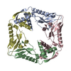
| Entry | Database: PDB / ID: 1hx5 | ||||||
|---|---|---|---|---|---|---|---|
| Title | Crystal structure of M. tuberculosis chaperonin-10 | ||||||
 Components Components | 10 KDA CHAPERONIN | ||||||
 Keywords Keywords | CHAPERONE / beta barrel / mobile loop / Structural Genomics / PSI / Protein Structure Initiative / TB Structural Genomics Consortium / TBSGC | ||||||
| Function / homology |  Function and homology information Function and homology information: / cell wall / zymogen binding / protein folding chaperone / peptidoglycan-based cell wall / : / cellular response to heat / response to heat / protein-folding chaperone binding / response to antibiotic ...: / cell wall / zymogen binding / protein folding chaperone / peptidoglycan-based cell wall / : / cellular response to heat / response to heat / protein-folding chaperone binding / response to antibiotic / regulation of DNA-templated transcription / ATP hydrolysis activity / extracellular region / ATP binding / metal ion binding / plasma membrane / cytosol Similarity search - Function | ||||||
| Biological species |   Mycobacterium tuberculosis (bacteria) Mycobacterium tuberculosis (bacteria) | ||||||
| Method |  X-RAY DIFFRACTION / MOLECULAR REPLACEMENT / Resolution: 3.5 Å X-RAY DIFFRACTION / MOLECULAR REPLACEMENT / Resolution: 3.5 Å | ||||||
 Authors Authors | Taneja, B. / Mande, S.C. / TB Structural Genomics Consortium (TBSGC) | ||||||
 Citation Citation | Journal: CURR.SCI. / Year: 2001 Title: Three-dimensional Structure of Mycobacterium tuberculosis Chaperonin-10 Reveals a Partially Stable Conformation for its Mobile Loop Authors: Taneja, B. / Mande, S.C. #1: Journal: Acta Crystallogr.,Sect.D / Year: 2002 Title: Structure of Mycobacterium tuberculosis chaperonin-10 at 3.5 A resolution. Authors: Taneja, B. / Mande, S.C. | ||||||
| History |
|
- Structure visualization
| Structure viewer | Molecule: MolmilJmol/JSmol |
|---|
- Downloads & links
-Download
| PDBx/mmCIF format | 1hx5.cif.gz | 103.6 KB | Display | PDBx/mmCIF format |
|---|---|---|---|---|
| PDB format | pdb1hx5.ent.gz | 85.2 KB | Display | PDB format |
| PDBx/mmJSON format | 1hx5.json.gz | Tree view | PDBx/mmJSON format | |
| Others |  Other downloads Other downloads |
-Validation report
| Arichive directory | https://data.pdbj.org/pub/pdb/validation_reports/hx/1hx5ftp://data.pdbj.org/pub/pdb/validation_reports/hx/1hx5 | HTTPS FTP |
|---|
-Related structure data
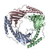
| Related structure data | 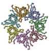 1lepS S: Starting model for refinement |
|---|---|
| Similar structure data | |
| Other databases |


-Links
 PDBj
PDBj

- Assembly
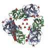
| Deposited unit | 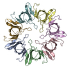
| ||||||||
|---|---|---|---|---|---|---|---|---|---|
| 1 |
| ||||||||
| Unit cell |
|
-Components
| #1: Protein | Mass: 10684.979 Da / Num. of mol.: 7 Source method: isolated from a genetically manipulated source Source: (gene. exp.) Mycobacterium tuberculosis (bacteria) / Gene: RV3418C / Plasmid: PMAL-C / Production host: Has protein modification | N | |
|---|
-Experimental details
-Experiment
| Experiment | Method: X-RAY DIFFRACTION / Number of used crystals: 1 |
|---|
- Sample preparation
| Crystal | Density Matthews: 2.64 Å3/Da / Density % sol: 53.47 % | ||||||||||||||||||||||||||||||||||||
|---|---|---|---|---|---|---|---|---|---|---|---|---|---|---|---|---|---|---|---|---|---|---|---|---|---|---|---|---|---|---|---|---|---|---|---|---|---|
| Crystal grow | Temperature: 298 K / Method: vapor diffusion, hanging drop / pH: 4 Details: PEG 400, lithium sulphate, sodium acetate, pH 4.0, VAPOR DIFFUSION, HANGING DROP, temperature 298K | ||||||||||||||||||||||||||||||||||||
| Crystal grow | *PLUS | ||||||||||||||||||||||||||||||||||||
| Components of the solutions | *PLUS
|
-Data collection
| Diffraction | Mean temperature: 298 K |
|---|---|
| Diffraction source | Source: ROTATING ANODE / Type: RIGAKU / Wavelength: 1.5418 Å |
| Detector | Type: MARRESEARCH / Detector: IMAGE PLATE / Date: Nov 15, 2000 / Details: mirrors |
| Radiation | Monochromator: mirrors / Protocol: SINGLE WAVELENGTH / Monochromatic (M) / Laue (L): M / Scattering type: x-ray |
| Radiation wavelength | Wavelength: 1.5418 Å / Relative weight: 1 |
| Reflection | Resolution: 3.5→30 Å / Num. all: 9954 / Num. obs: 9927 / % possible obs: 96.4 % / Observed criterion σ(F): 0 / Observed criterion σ(I): 0 / Redundancy: 7.7 % / Biso Wilson estimate: 68.6 Å2 / Rmerge(I) obs: 0.078 / Net I/σ(I): 11.7 |
| Reflection shell | Resolution: 3.5→3.77 Å / Redundancy: 3.5 % / Rmerge(I) obs: 0.28 / % possible all: 90.5 |
| Reflection | *PLUS Lowest resolution: 30 Å |
- Processing
| Software |
| |||||||||||||||||||||||||
|---|---|---|---|---|---|---|---|---|---|---|---|---|---|---|---|---|---|---|---|---|---|---|---|---|---|---|
| Refinement | Method to determine structure: MOLECULAR REPLACEMENT Starting model: PDB entry 1LEP Resolution: 3.5→30 Å / σ(F): 0 / σ(I): 0 / Stereochemistry target values: Engh and Huber
| |||||||||||||||||||||||||
| Refinement step | Cycle: LAST / Resolution: 3.5→30 Å
| |||||||||||||||||||||||||
| Software | *PLUS Name: CNS / Classification: refinement | |||||||||||||||||||||||||
| Refinement | *PLUS Lowest resolution: 30 Å / σ(F): 0 | |||||||||||||||||||||||||
| Solvent computation | *PLUS | |||||||||||||||||||||||||
| Displacement parameters | *PLUS | |||||||||||||||||||||||||
| Refine LS restraints | *PLUS
|
