 Movie
Movie Controller


+ Open data

- Basic information
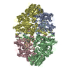
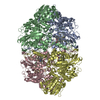
| Entry | Database: PDB / ID: 1qf7 | ||||||
|---|---|---|---|---|---|---|---|
| Title | STRUCTURE OF THE MUTANT HIS392GLN OF CATALASE HPII FROM E. COLI | ||||||
 Components Components | PROTEIN (CATALASE HPII) | ||||||
 Keywords Keywords | OXIDOREDUCTASE / COVALENT MODIFICATIONS | ||||||
| Function / homology |  Function and homology information Function and homology informationcatalase / catalase activity / hyperosmotic response / hydrogen peroxide catabolic process / response to oxidative stress / iron ion binding / heme binding / DNA damage response / identical protein binding / cytoplasm / cytosol Similarity search - Function | ||||||
| Biological species |  | ||||||
| Method |  X-RAY DIFFRACTION / OTHER / Resolution: 2.2 Å X-RAY DIFFRACTION / OTHER / Resolution: 2.2 Å | ||||||
 Authors Authors | Mate, M.J. / Loewen, P.C. / Fita, I. | ||||||
 Citation Citation | Journal: J.Biol.Chem. / Year: 1999 Title: Mutants that alter the covalent structure of catalase hydroperoxidase II from Escherichia coli. Authors: Mate, M.J. / Sevinc, M.S. / Hu, B. / Bujons, J. / Bravo, J. / Switala, J. / Ens, W. / Loewen, P.C. / Fita, I. #1: Journal: Proteins / Year: 1999 Title: Structure of catalase HPII from Escherichia coli at 1.9 A resolution. Authors: Bravo, J. / Mate, M.J. / Schneider, T. / Switala, J. / Wilson, K. / Loewen, P.C. / Fita, I. | ||||||
| History |
|
- Structure visualization
| Structure viewer | Molecule: MolmilJmol/JSmol |
|---|
- Downloads & links
-Download
| PDBx/mmCIF format | 1qf7.cif.gz | 631.7 KB | Display | PDBx/mmCIF format |
|---|---|---|---|---|
| PDB format | pdb1qf7.ent.gz | 510.4 KB | Display | PDB format |
| PDBx/mmJSON format | 1qf7.json.gz | Tree view | PDBx/mmJSON format | |
| Others |  Other downloads Other downloads |
-Validation report
| Arichive directory | https://data.pdbj.org/pub/pdb/validation_reports/qf/1qf7ftp://data.pdbj.org/pub/pdb/validation_reports/qf/1qf7 | HTTPS FTP |
|---|
-Related structure data
| Related structure data |  1cf9C  1iphS S: Starting model for refinement C: citing same article ( |
|---|---|
| Similar structure data |


-Links
 PDBj
PDBj

- Assembly
| Deposited unit | 
| ||||||||
|---|---|---|---|---|---|---|---|---|---|
| 1 |
| ||||||||
| Unit cell |
|
-Components
| #1: Protein | Mass: 84261.438 Da / Num. of mol.: 4 / Mutation: H392Q Source method: isolated from a genetically manipulated source Source: (gene. exp.) #2: Chemical | ChemComp-HEM /   Mass: 616.487 Da / Num. of mol.: 4 / Source method: obtained synthetically / Formula: C34H32FeN4O4 Mass: 616.487 Da / Num. of mol.: 4 / Source method: obtained synthetically / Formula: C34H32FeN4O4#3: Water | ChemComp-HOH / |  Mass: 18.015 Da / Num. of mol.: 2679 / Source method: isolated from a natural source / Formula: H2O Mass: 18.015 Da / Num. of mol.: 2679 / Source method: isolated from a natural source / Formula: H2O |
|---|
-Experimental details
-Experiment
| Experiment | Method: X-RAY DIFFRACTION / Number of used crystals: 1 |
|---|
- Sample preparation
| Crystal | Density Matthews: 2.11 Å3/Da / Density % sol: 41.74 % | ||||||||||||||||||||||||
|---|---|---|---|---|---|---|---|---|---|---|---|---|---|---|---|---|---|---|---|---|---|---|---|---|---|
| Crystal grow | pH: 9 / Details: pH 9 | ||||||||||||||||||||||||
| Crystal grow | *PLUS pH: 9 / Method: vapor diffusion, hanging drop / Details: Bravo, J., (1995) Structure, 3, 491. | ||||||||||||||||||||||||
| Components of the solutions | *PLUS
|
-Data collection
| Diffraction | Mean temperature: 100 K |
|---|---|
| Diffraction source | Source: ROTATING ANODE / Type: RIGAKU RU200 / Wavelength: 1.5418 |
| Detector | Detector: IMAGE PLATE / Date: Oct 1, 1998 |
| Radiation | Monochromator: NI FILTER / Protocol: SINGLE WAVELENGTH / Monochromatic (M) / Laue (L): M / Scattering type: x-ray |
| Radiation wavelength | Wavelength: 1.5418 Å / Relative weight: 1 |
| Reflection | Resolution: 2.12→20 Å / Num. obs: 137604 / % possible obs: 96.6 % / Observed criterion σ(I): 2 / Redundancy: 3 % / Biso Wilson estimate: 13.63 Å2 / Rmerge(I) obs: 0.08 / Rsym value: 0.09 / Net I/σ(I): 10.4 |
| Reflection shell | Resolution: 2.12→2.2 Å / Redundancy: 2.5 % / Rmerge(I) obs: 0.21 / Mean I/σ(I) obs: 5.1 / Rsym value: 0.25 / % possible all: 95 |
| Reflection shell | *PLUS % possible obs: 95 % |
- Processing
| Software |
| ||||||||||||||||||||||||||||||||||||||||||||||||||||||||||||||||||||||||||||||||||||
|---|---|---|---|---|---|---|---|---|---|---|---|---|---|---|---|---|---|---|---|---|---|---|---|---|---|---|---|---|---|---|---|---|---|---|---|---|---|---|---|---|---|---|---|---|---|---|---|---|---|---|---|---|---|---|---|---|---|---|---|---|---|---|---|---|---|---|---|---|---|---|---|---|---|---|---|---|---|---|---|---|---|---|---|---|---|
| Refinement | Method to determine structure: OTHER Starting model: PDB ENTRY 1IPH Resolution: 2.2→20 Å / SU B: 5.565 / SU ML: 0.141 / Cross valid method: THROUGHOUT / σ(F): 0 / ESU R: 0.318 / ESU R Free: 0.211
| ||||||||||||||||||||||||||||||||||||||||||||||||||||||||||||||||||||||||||||||||||||
| Displacement parameters | Biso mean: 13.55 Å2
| ||||||||||||||||||||||||||||||||||||||||||||||||||||||||||||||||||||||||||||||||||||
| Refinement step | Cycle: LAST / Resolution: 2.2→20 Å
| ||||||||||||||||||||||||||||||||||||||||||||||||||||||||||||||||||||||||||||||||||||
| Refine LS restraints |
| ||||||||||||||||||||||||||||||||||||||||||||||||||||||||||||||||||||||||||||||||||||
| Software | *PLUS Name: REFMAC / Classification: refinement | ||||||||||||||||||||||||||||||||||||||||||||||||||||||||||||||||||||||||||||||||||||
| Refinement | *PLUS Highest resolution: 2.2 Å / σ(F): 0 / % reflection Rfree: 5 % / Rfactor obs: 0.144 / Rfactor Rfree: 0.21 | ||||||||||||||||||||||||||||||||||||||||||||||||||||||||||||||||||||||||||||||||||||
| Solvent computation | *PLUS | ||||||||||||||||||||||||||||||||||||||||||||||||||||||||||||||||||||||||||||||||||||
| Displacement parameters | *PLUS | ||||||||||||||||||||||||||||||||||||||||||||||||||||||||||||||||||||||||||||||||||||
| Refine LS restraints | *PLUS
|
