 Movie
Movie Controller


+ Open data

- Basic information
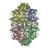
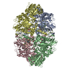
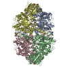
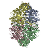
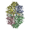
| Entry | Database: PDB / ID: 1iph | |||||||||
|---|---|---|---|---|---|---|---|---|---|---|
| Title | STRUCTURE OF CATALASE HPII FROM ESCHERICHIA COLI | |||||||||
 Components Components | CATALASE HPII | |||||||||
 Keywords Keywords | OXIDOREDUCTASE / HYDROGEN PEROXIDE | |||||||||
| Function / homology |  Function and homology information Function and homology informationcatalase / catalase activity / hyperosmotic response / hydrogen peroxide catabolic process / response to oxidative stress / iron ion binding / heme binding / DNA damage response / identical protein binding / cytoplasm / cytosol Similarity search - Function | |||||||||
| Biological species |  | |||||||||
| Method |  X-RAY DIFFRACTION / Resolution: 2.8 Å X-RAY DIFFRACTION / Resolution: 2.8 Å | |||||||||
 Authors Authors | Bravo, J. / Loewen, P.C. / Fita, I. | |||||||||
 Citation Citation | Journal: Structure / Year: 1995 Title: Crystal structure of catalase HPII from Escherichia coli. Authors: Bravo, J. / Verdaguer, N. / Tormo, J. / Betzel, C. / Switala, J. / Loewen, P.C. / Fita, I. #1: Journal: Joint Ccp4 Esf-Eacbm Newsletter on Protein CrystallographyYear: 1993 Title: 2.8 A Crystal Structure of Catalase Hpii from Escherichia Coli Authors: Bravo, J. / Verdaguer, N. / Tormo, J. / Betzel, C. / Switala, J. / Loewen, P.C. / Fita, I. #2: Journal: J.Mol.Biol. / Year: 1990Title: Crystallization and Preliminary X-Ray Diffraction Analysis of Catalase Hpii from Escherichia Coli Authors: Tormo, J. / Fita, I. / Switala, J. / Loewen, P.C. #3: Journal: Acta Crystallogr.,Sect.B / Year: 1986Title: The Refined Structure of Beef Liver Catalase at 2.5 A Resolution Authors: Fita, I. / Silva, A.M. / Murthy, M.R.N. / Rossmann, M.G. | |||||||||
| History |
|
- Structure visualization
| Structure viewer | Molecule: MolmilJmol/JSmol |
|---|
- Downloads & links
-Download
| PDBx/mmCIF format | 1iph.cif.gz | 544 KB | Display | PDBx/mmCIF format |
|---|---|---|---|---|
| PDB format | pdb1iph.ent.gz | 443 KB | Display | PDB format |
| PDBx/mmJSON format | 1iph.json.gz | Tree view | PDBx/mmJSON format | |
| Others |  Other downloads Other downloads |
-Validation report
| Arichive directory | https://data.pdbj.org/pub/pdb/validation_reports/ip/1iphftp://data.pdbj.org/pub/pdb/validation_reports/ip/1iph | HTTPS FTP |
|---|
-Related structure data
| Similar structure data |
|---|


-Links
 PDBj
PDBj

- Assembly
| Deposited unit | 
| ||||||||||||||||
|---|---|---|---|---|---|---|---|---|---|---|---|---|---|---|---|---|---|
| 1 |
| ||||||||||||||||
| Unit cell |
| ||||||||||||||||
| Noncrystallographic symmetry (NCS) | NCS oper:
|
-Components
| #1: Protein | Mass: 84271.453 Da / Num. of mol.: 4 / Source method: isolated from a natural source / Source: (natural) #2: Chemical | ChemComp-HEM /   Mass: 616.487 Da / Num. of mol.: 4 / Source method: obtained synthetically / Formula: C34H32FeN4O4 Mass: 616.487 Da / Num. of mol.: 4 / Source method: obtained synthetically / Formula: C34H32FeN4O4 |
|---|
-Experimental details
-Experiment
| Experiment | Method: X-RAY DIFFRACTION |
|---|
- Sample preparation
| Crystal | Density Matthews: 2.23 Å3/Da / Density % sol: 44.85 % | ||||||||||||||||||||||||
|---|---|---|---|---|---|---|---|---|---|---|---|---|---|---|---|---|---|---|---|---|---|---|---|---|---|
| Crystal grow | *PLUS pH: 9 / Method: vapor diffusion, hanging drop | ||||||||||||||||||||||||
| Components of the solutions | *PLUS
|
-Data collection
| Radiation | Scattering type: x-ray |
|---|---|
| Radiation wavelength | Relative weight: 1 |
| Reflection | *PLUS Highest resolution: 2.8 Å / Num. obs: 225245 / % possible obs: 90 % / Rmerge(I) obs: 0.108 / Num. measured all: 65329 |
| Reflection shell | *PLUS Mean I/σ(I) obs: 3.5 |
- Processing
| Software |
| ||||||||||||||||||||||||||||||||||||||||||||||||||||||||||||
|---|---|---|---|---|---|---|---|---|---|---|---|---|---|---|---|---|---|---|---|---|---|---|---|---|---|---|---|---|---|---|---|---|---|---|---|---|---|---|---|---|---|---|---|---|---|---|---|---|---|---|---|---|---|---|---|---|---|---|---|---|---|
| Refinement | Rfactor Rwork: 0.2 / Rfactor obs: 0.2 / Highest resolution: 2.8 Å | ||||||||||||||||||||||||||||||||||||||||||||||||||||||||||||
| Refinement step | Cycle: LAST / Highest resolution: 2.8 Å
| ||||||||||||||||||||||||||||||||||||||||||||||||||||||||||||
| Refine LS restraints |
| ||||||||||||||||||||||||||||||||||||||||||||||||||||||||||||
| Refinement | *PLUS Lowest resolution: 8 Å / Num. reflection obs: 58477 | ||||||||||||||||||||||||||||||||||||||||||||||||||||||||||||
| Solvent computation | *PLUS | ||||||||||||||||||||||||||||||||||||||||||||||||||||||||||||
| Displacement parameters | *PLUS |
