 Movie
Movie Controller

[English] 日本語
 Yorodumi
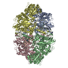
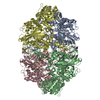
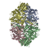
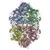
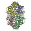
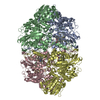
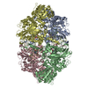
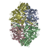
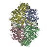
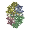
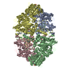
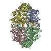
- PDB-1ggf: CRYSTAL STRUCTURE OF CATALASE HPII FROM ESCHERICHIA COLI, VARIANT... -
+ Open data

- Basic information
| Entry | Database: PDB / ID: 1ggf | ||||||
|---|---|---|---|---|---|---|---|
| Title | CRYSTAL STRUCTURE OF CATALASE HPII FROM ESCHERICHIA COLI, VARIANT HIS128ASN, COMPLEX WITH HYDROGEN PEROXIDE. | ||||||
 Components Components | CATALASE HPII | ||||||
 Keywords Keywords | OXIDOREDUCTASE / BETA BARREL / ALPHA HELICAL DOMAIN / FLAVODOXIN LIKE DOMAIN | ||||||
| Function / homology |  Function and homology information Function and homology informationcatalase / hyperosmotic response / catalase activity / hydrogen peroxide catabolic process / response to oxidative stress / iron ion binding / heme binding / DNA damage response / identical protein binding / cytosol / cytoplasm Similarity search - Function | ||||||
| Biological species |  | ||||||
| Method |  X-RAY DIFFRACTION / Resolution: 2.28 Å X-RAY DIFFRACTION / Resolution: 2.28 Å | ||||||
 Authors Authors | Melik-Adamyan, W.R. / Bravo, J. / Carpena, X. / Switala, J. / Mate, M.J. / Fita, I. / Loewen, P.C. | ||||||
 Citation Citation | Journal: Proteins / Year: 2001 Title: Substrate flow in catalases deduced from the crystal structures of active site variants of HPII from Escherichia coli. Authors: Melik-Adamyan, W. / Bravo, J. / Carpena, X. / Switala, J. / Mate, M.J. / Fita, I. / Loewen, P.C. #1: Journal: Structure / Year: 1995Title: Crystal Structure of Catalase HPII from Escherichia coli Authors: Bravo, J. / Verdaguer, N. / Tormo, J. / Betzel, C. / Switala, J. / Loewen, P.C. / Fita, I. #2: Journal: PROTEINS: STRUCT.,FUNCT.,GENET. / Year: 1999Title: Structure of Catalase Hpii from Escherichia Coli at 1.9 A Resolution Authors: Bravo, J. / Mate, M.J. / Schneider, T. / Switala, J. / Wilson, K. / Loewen, P.C. / Fita, I. | ||||||
| History |
|
- Structure visualization
| Structure viewer | Molecule: MolmilJmol/JSmol |
|---|
- Downloads & links
-Download
| PDBx/mmCIF format | 1ggf.cif.gz | 613.6 KB | Display | PDBx/mmCIF format |
|---|---|---|---|---|
| PDB format | pdb1ggf.ent.gz | 498.6 KB | Display | PDB format |
| PDBx/mmJSON format | 1ggf.json.gz | Tree view | PDBx/mmJSON format | |
| Others |  Other downloads Other downloads |
-Validation report
| Arichive directory | https://data.pdbj.org/pub/pdb/validation_reports/gg/1ggfftp://data.pdbj.org/pub/pdb/validation_reports/gg/1ggf | HTTPS FTP |
|---|
-Related structure data


-Links
 PDBj
PDBj

- Assembly
| Deposited unit | 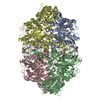
| ||||||||
|---|---|---|---|---|---|---|---|---|---|
| 1 |
| ||||||||
| Unit cell |
|
-Components
| #1: Protein | Mass: 84247.406 Da / Num. of mol.: 4 / Mutation: H128N Source method: isolated from a genetically manipulated source Source: (gene. exp.) #2: Chemical | ChemComp-HEM /   Mass: 616.487 Da / Num. of mol.: 4 / Source method: obtained synthetically / Formula: C34H32FeN4O4 Mass: 616.487 Da / Num. of mol.: 4 / Source method: obtained synthetically / Formula: C34H32FeN4O4#3: Chemical | ChemComp-PEO / 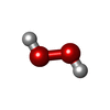  Mass: 34.015 Da / Num. of mol.: 13 / Source method: obtained synthetically / Formula: H2O2 Mass: 34.015 Da / Num. of mol.: 13 / Source method: obtained synthetically / Formula: H2O2#4: Water | ChemComp-HOH / |  Mass: 18.015 Da / Num. of mol.: 2025 / Source method: isolated from a natural source / Formula: H2O Mass: 18.015 Da / Num. of mol.: 2025 / Source method: isolated from a natural source / Formula: H2O |
|---|
-Experimental details
-Experiment
| Experiment | Method: X-RAY DIFFRACTION |
|---|
- Sample preparation
| Crystal | Density Matthews: 2.12 Å3/Da / Density % sol: 42.01 % | ||||||||||||||||||||||||||||||
|---|---|---|---|---|---|---|---|---|---|---|---|---|---|---|---|---|---|---|---|---|---|---|---|---|---|---|---|---|---|---|---|
| Crystal grow | Temperature: 297 K / pH: 9 Details: PEG 3350, LiCl, Tris-HCl. Before data collection the protein crystal was soaked during a few seconds in the solution with 2M hydrogen peroxide., pH 9.0, temperature 297.0K | ||||||||||||||||||||||||||||||
| Crystal grow | *PLUS pH: 9 / Method: vapor diffusion, hanging drop | ||||||||||||||||||||||||||||||
| Components of the solutions | *PLUS
|
-Data collection
| Diffraction | Mean temperature: 120 K |
|---|---|
| Detector | Type: MARRESEARCH / Detector: IMAGE PLATE / Date: Jan 15, 1996 |
| Radiation | Protocol: SINGLE WAVELENGTH / Monochromatic (M) / Laue (L): M / Scattering type: x-ray |
| Radiation wavelength | Relative weight: 1 |
| Reflection | Resolution: 2.28→19.84 Å / Num. all: 128033 / Num. obs: 125885 / % possible obs: 98.5 % / Biso Wilson estimate: 33.9 Å2 |
| Reflection shell | Resolution: 2.28→2.34 Å / Num. unique all: 9488 / % possible all: 95.9 |
| Reflection | *PLUS Num. all: 128033 / Rmerge(I) obs: 0.107 |
| Reflection shell | *PLUS % possible obs: 95.9 % / Num. unique obs: 9488 / Rmerge(I) obs: 0.365 |
- Processing
| Software |
| ||||||||||||||||||||
|---|---|---|---|---|---|---|---|---|---|---|---|---|---|---|---|---|---|---|---|---|---|
| Refinement | Resolution: 2.28→87.95 Å / Stereochemistry target values: CCP4, protin_jp.idl Details: REFMAC, WEIGHT MATRIX 0.2. X-Plor was also used during refinement.
| ||||||||||||||||||||
| Refinement step | Cycle: LAST / Resolution: 2.28→87.95 Å
| ||||||||||||||||||||
| Refine LS restraints |
| ||||||||||||||||||||
| Software | *PLUS Name: REFMAC / Classification: refinement | ||||||||||||||||||||
| Refinement | *PLUS Lowest resolution: 19.8 Å | ||||||||||||||||||||
| Solvent computation | *PLUS | ||||||||||||||||||||
| Displacement parameters | *PLUS | ||||||||||||||||||||
| Refine LS restraints | *PLUS
| ||||||||||||||||||||
| LS refinement shell | *PLUS Highest resolution: 2.28 Å / Lowest resolution: 2.34 Å / Rfactor Rfree: 0.296 / Rfactor obs: 0.198 |
