 ムービー
ムービー コントローラー


+ データを開く

- 基本情報
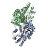
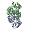
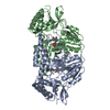
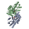
基本情報
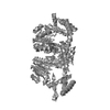
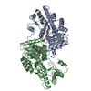
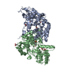
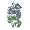
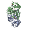
| 登録情報 | データベース: PDB / ID: 1p3w | ||||||
|---|---|---|---|---|---|---|---|
| タイトル | X-ray crystal structure of E. coli IscS | ||||||
 要素 要素 | Cysteine desulfurase | ||||||
 キーワード キーワード | LYASE / iron sulfur cluster / Nifs / CsdB | ||||||
| 機能・相同性 |  機能・相同性情報 機能・相同性情報: / IscS-TusA complex / IscS-IscU complex / tRNA 4-thiouridine biosynthesis / sulfur compound transport / selenocysteine catabolic process / sulfurtransferase complex / detection of UV / selenocysteine lyase activity / tRNA wobble position uridine thiolation ...: / IscS-TusA complex / IscS-IscU complex / tRNA 4-thiouridine biosynthesis / sulfur compound transport / selenocysteine catabolic process / sulfurtransferase complex / detection of UV / selenocysteine lyase activity / tRNA wobble position uridine thiolation / L-cysteine desulfurase complex / sulfur carrier activity / L-cysteine catabolic process / cysteine desulfurase / cysteine desulfurase activity / thiamine biosynthetic process / [2Fe-2S] cluster assembly / iron-sulfur cluster assembly / 2 iron, 2 sulfur cluster binding / pyridoxal phosphate binding / metal ion binding / cytosol 類似検索 - 分子機能 | ||||||
| 生物種 |  | ||||||
| 手法 |  X線回折 / シンクロトロン / 分子置換 / 解像度: 2.1 Å X線回折 / シンクロトロン / 分子置換 / 解像度: 2.1 Å | ||||||
 データ登録者 データ登録者 | Cupp-Vickery, J.R. / Vickery, L.E. / Urbina, H. | ||||||
 引用 引用 | ジャーナル: J.Mol.Biol. / 年: 2003 タイトル: Crystal Structure of IscS, a Cysteine Desulfurase from Escherichia coli 著者: Cupp-Vickery, J.R. / Urbina, H. / Vickery, L.E. | ||||||
| 履歴 |
|
- 構造の表示
構造の表示
| 構造ビューア | 分子: MolmilJmol/JSmol |
|---|
- ダウンロードとリンク
-ダウンロード
| PDBx/mmCIF形式 | 1p3w.cif.gz | 173.3 KB | 表示 | PDBx/mmCIF形式 |
|---|---|---|---|---|
| PDB形式 | pdb1p3w.ent.gz | 136.5 KB | 表示 | PDB形式 |
| PDBx/mmJSON形式 | 1p3w.json.gz | ツリー表示 | PDBx/mmJSON形式 | |
| その他 |  その他のダウンロード その他のダウンロード |
-検証レポート
| アーカイブディレクトリ | https://data.pdbj.org/pub/pdb/validation_reports/p3/1p3wftp://data.pdbj.org/pub/pdb/validation_reports/p3/1p3w | HTTPS FTP |
|---|
-関連構造データ

-リンク
 PDBj
PDBj- 集合体
集合体
| 登録構造単位 | 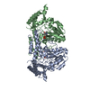
| ||||||||
|---|---|---|---|---|---|---|---|---|---|
| 1 |
| ||||||||
| 単位格子 |
| ||||||||
| 詳細 | Biological unit is a dimer. |
-要素
| #1: タンパク質 | 分子量: 45149.477 Da / 分子数: 2 / 由来タイプ: 組換発現 / 由来: (組換発現) 遺伝子: ISCS OR B2530 OR C3056 OR Z3797 OR ECS3396 OR SF2577 発現宿主: 参照: UniProt: P0A6B7, 付加脱離酵素(リアーゼ); C-Sリアーゼ類; - #2: 化合物 |   分子量: 247.142 Da / 分子数: 2 / 由来タイプ: 合成 / 式: C8H10NO6P 分子量: 247.142 Da / 分子数: 2 / 由来タイプ: 合成 / 式: C8H10NO6P#3: 水 | ChemComp-HOH / |  分子量: 18.015 Da / 分子数: 625 / 由来タイプ: 天然 / 式: H2O 分子量: 18.015 Da / 分子数: 625 / 由来タイプ: 天然 / 式: H2O |
|---|
-実験情報
-実験
| 実験 | 手法: X線回折 / 使用した結晶の数: 1 |
|---|
- 試料調製
試料調製
| 結晶 | マシュー密度: 2.19 Å3/Da / 溶媒含有率: 43.51 % | ||||||||||||||||||||||||||||||
|---|---|---|---|---|---|---|---|---|---|---|---|---|---|---|---|---|---|---|---|---|---|---|---|---|---|---|---|---|---|---|---|
| 結晶化 | 温度: 295.1 K / 手法: 蒸気拡散法, ハンギングドロップ法 / pH: 7.5 詳細: PEG, pH 7.5, VAPOR DIFFUSION, HANGING DROP, temperature 22.1K | ||||||||||||||||||||||||||||||
| 結晶化 | *PLUS pH: 6.5 / 手法: 蒸気拡散法, ハンギングドロップ法 | ||||||||||||||||||||||||||||||
| 溶液の組成 | *PLUS
|
-データ収集
| 回折 | 平均測定温度: 103 K |
|---|---|
| 放射光源 | 由来: シンクロトロン / サイト: SSRL  / ビームライン: BL9-2 / ビームライン: BL9-2 |
| 検出器 | タイプ: ADSC QUANTUM 4 / 検出器: CCD |
| 放射 | プロトコル: SINGLE WAVELENGTH / 単色(M)・ラウエ(L): M / 散乱光タイプ: x-ray |
| 放射波長 | 相対比: 1 |
| 反射 | 解像度: 2.1→50 Å / Num. all: 46748 / Num. obs: 46748 / % possible obs: 97 % / Observed criterion σ(F): 0 / Observed criterion σ(I): 0 / 冗長度: 3.2 % / Rsym value: 0.068 / Net I/σ(I): 7.1 |
| 反射 シェル | 解像度: 2.1→2.27 Å / 冗長度: 3.1 % / Mean I/σ(I) obs: 2.7 / Num. unique all: 3435 / Rsym value: 0.245 / % possible all: 93.3 |
| 反射 | *PLUS 最低解像度: 500 Å / % possible obs: 96.9 % / Num. measured all: 146645 / Rmerge(I) obs: 0.068 |
| 反射 シェル | *PLUS 最低解像度: 2.15 Å / % possible obs: 93.3 % / Rmerge(I) obs: 0.245 |
- 解析
解析
| ソフトウェア |
| ||||||||||||||||||||
|---|---|---|---|---|---|---|---|---|---|---|---|---|---|---|---|---|---|---|---|---|---|
| 精密化 | 構造決定の手法: 分子置換 開始モデル: T.m. Nifs 解像度: 2.1→5 Å / σ(F): 0 / σ(I): 0 / 立体化学のターゲット値: Engh & Huber
| ||||||||||||||||||||
| 精密化ステップ | サイクル: LAST / 解像度: 2.1→5 Å
| ||||||||||||||||||||
| LS精密化 シェル | 解像度: 2.1→2.11 Å
| ||||||||||||||||||||
| 拘束条件 | *PLUS
| ||||||||||||||||||||
| LS精密化 シェル | *PLUS 最高解像度: 2.1 Å |
