 Movie
Movie Controller


+ Open data

- Basic information
| Entry | Database: PDB / ID: 1ay4 | ||||||
|---|---|---|---|---|---|---|---|
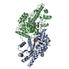
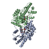
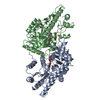
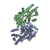
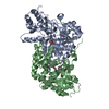
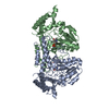
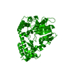
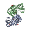
| Title | AROMATIC AMINO ACID AMINOTRANSFERASE WITHOUT SUBSTRATE | ||||||
 Components Components | AROMATIC AMINO ACID AMINOTRANSFERASE | ||||||
 Keywords Keywords | TRANSFERASE / AMINOTRANSFERASE | ||||||
| Function / homology |  Function and homology information Function and homology informationaromatic-amino-acid transaminase / : / L-tyrosine:2-oxoglutarate transaminase activity / L-aspartate:2-oxoglutarate transaminase activity / pyridoxal phosphate binding / identical protein binding / cytosol Similarity search - Function | ||||||
| Biological species |  Paracoccus denitrificans (bacteria) Paracoccus denitrificans (bacteria) | ||||||
| Method |  X-RAY DIFFRACTION / MOLECULAR REPLACEMENT / Resolution: 2.33 Å X-RAY DIFFRACTION / MOLECULAR REPLACEMENT / Resolution: 2.33 Å | ||||||
 Authors Authors | Okamoto, A. / Hirotsu, K. / Kagamiyama, H. | ||||||
 Citation Citation | Journal: J.Mol.Biol. / Year: 1998 Title: Crystal structures of Paracoccus denitrificans aromatic amino acid aminotransferase: a substrate recognition site constructed by rearrangement of hydrogen bond network. Authors: Okamoto, A. / Nakai, Y. / Hayashi, H. / Hirotsu, K. / Kagamiyama, H. #1: Journal: J.Biochem.(Tokyo) / Year: 1997Title: Paracoccus Denitrificans Aromatic Amino Acid Aminotransferase: A Model Enzyme for the Study of Dual Substrate Recognition Mechanism Authors: Oue, S. / Okamoto, A. / Nakai, Y. / Nakahira, M. / Shibatani, T. / Hayashi, H. / Kagamiyama, H. | ||||||
| History |
|
- Structure visualization
| Structure viewer | Molecule: MolmilJmol/JSmol |
|---|
- Downloads & links
-Download
| PDBx/mmCIF format | 1ay4.cif.gz | 159.8 KB | Display | PDBx/mmCIF format |
|---|---|---|---|---|
| PDB format | pdb1ay4.ent.gz | 129.6 KB | Display | PDB format |
| PDBx/mmJSON format | 1ay4.json.gz | Tree view | PDBx/mmJSON format | |
| Others |  Other downloads Other downloads |
-Validation report
| Arichive directory | https://data.pdbj.org/pub/pdb/validation_reports/ay/1ay4ftp://data.pdbj.org/pub/pdb/validation_reports/ay/1ay4 | HTTPS FTP |
|---|
-Related structure data
| Related structure data |  1ay5C 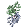 1ay8C 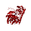 1artS S: Starting model for refinement C: citing same article ( |
|---|---|
| Similar structure data |


-Links
 PDBj
PDBj- Assembly
| Deposited unit | 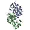
| ||||||||
|---|---|---|---|---|---|---|---|---|---|
| 1 |
| ||||||||
| Unit cell |
| ||||||||
| Noncrystallographic symmetry (NCS) | NCS oper: (Code: given Matrix: (-0.95702, 0.122115, -0.263058), Vector: |
-Components
| #1: Protein | Mass: 42779.996 Da / Num. of mol.: 2 Source method: isolated from a genetically manipulated source Source: (gene. exp.) Paracoccus denitrificans (bacteria) / Strain: IFO12442 / Plasmid: PUC118 / Production host: References: UniProt: P95468, aromatic-amino-acid transaminase #2: Chemical |   Mass: 247.142 Da / Num. of mol.: 2 / Source method: obtained synthetically / Formula: C8H10NO6P Mass: 247.142 Da / Num. of mol.: 2 / Source method: obtained synthetically / Formula: C8H10NO6P#3: Water | ChemComp-HOH / |  Mass: 18.015 Da / Num. of mol.: 321 / Source method: isolated from a natural source / Formula: H2O Mass: 18.015 Da / Num. of mol.: 321 / Source method: isolated from a natural source / Formula: H2O |
|---|
-Experimental details
-Experiment
| Experiment | Method: X-RAY DIFFRACTION / Number of used crystals: 1 |
|---|
- Sample preparation
| Crystal | Density Matthews: 2.4 Å3/Da / Density % sol: 48.8 % | ||||||||||||||||||||||||||||||||||||||||||||||||||||||
|---|---|---|---|---|---|---|---|---|---|---|---|---|---|---|---|---|---|---|---|---|---|---|---|---|---|---|---|---|---|---|---|---|---|---|---|---|---|---|---|---|---|---|---|---|---|---|---|---|---|---|---|---|---|---|---|
| Crystal grow | pH: 5.7 Details: PROTEIN WAS CRYSTALLIZED FROM 16.5% PEG 4000, 0.4 M SODIUM ACETATE, PH 5.7 | ||||||||||||||||||||||||||||||||||||||||||||||||||||||
| Crystal | *PLUS | ||||||||||||||||||||||||||||||||||||||||||||||||||||||
| Crystal grow | *PLUS Temperature: 20 ℃ / Method: vapor diffusion, sitting drop / Details: used to seeding | ||||||||||||||||||||||||||||||||||||||||||||||||||||||
| Components of the solutions | *PLUS
|
-Data collection
| Diffraction | Mean temperature: 293 K |
|---|---|
| Diffraction source | Source: ROTATING ANODE / Type: RIGAKU RUH2R / Wavelength: 1.5418 |
| Detector | Type: RIGAKU / Detector: IMAGE PLATE / Date: Mar 7, 1996 / Details: MIRRORS |
| Radiation | Monochromator: NI FILTER / Monochromatic (M) / Laue (L): M / Scattering type: x-ray |
| Radiation wavelength | Wavelength: 1.5418 Å / Relative weight: 1 |
| Reflection | Resolution: 2.33→123 Å / Num. obs: 33010 / % possible obs: 97.4 % / Observed criterion σ(I): 1 / Redundancy: 3.4 % / Biso Wilson estimate: 18.4 Å2 / Rmerge(I) obs: 0.0752 / Net I/σ(I): 11.5 |
| Reflection shell | Resolution: 2.33→2.5 Å / Redundancy: 2.4 % / Rmerge(I) obs: 0.174 / Mean I/σ(I) obs: 4 / % possible all: 95.4 |
| Reflection | *PLUS Num. measured all: 110551 |
- Processing
| Software |
| ||||||||||||||||||||||||||||||||||||||||||||||||||||||||||||||||||||||||||||||||
|---|---|---|---|---|---|---|---|---|---|---|---|---|---|---|---|---|---|---|---|---|---|---|---|---|---|---|---|---|---|---|---|---|---|---|---|---|---|---|---|---|---|---|---|---|---|---|---|---|---|---|---|---|---|---|---|---|---|---|---|---|---|---|---|---|---|---|---|---|---|---|---|---|---|---|---|---|---|---|---|---|---|
| Refinement | Method to determine structure: MOLECULAR REPLACEMENT Starting model: 1ART Resolution: 2.33→6 Å / Rfactor Rfree error: 0.004 / Data cutoff high absF: 10000000 / Data cutoff low absF: 0.001 / Isotropic thermal model: RESTRAINED / Cross valid method: THROUGHOUT / σ(F): 2
| ||||||||||||||||||||||||||||||||||||||||||||||||||||||||||||||||||||||||||||||||
| Displacement parameters | Biso mean: 27.4 Å2 | ||||||||||||||||||||||||||||||||||||||||||||||||||||||||||||||||||||||||||||||||
| Refine analyze |
| ||||||||||||||||||||||||||||||||||||||||||||||||||||||||||||||||||||||||||||||||
| Refinement step | Cycle: LAST / Resolution: 2.33→6 Å
| ||||||||||||||||||||||||||||||||||||||||||||||||||||||||||||||||||||||||||||||||
| Refine LS restraints |
| ||||||||||||||||||||||||||||||||||||||||||||||||||||||||||||||||||||||||||||||||
| LS refinement shell | Resolution: 2.33→2.47 Å / Rfactor Rfree error: 0.013 / Total num. of bins used: 6
| ||||||||||||||||||||||||||||||||||||||||||||||||||||||||||||||||||||||||||||||||
| Xplor file |
| ||||||||||||||||||||||||||||||||||||||||||||||||||||||||||||||||||||||||||||||||
| Software | *PLUS Name: X-PLOR / Version: 3.1 / Classification: refinement | ||||||||||||||||||||||||||||||||||||||||||||||||||||||||||||||||||||||||||||||||
| Refine LS restraints | *PLUS
|
