 Movie
Movie Controller

[English] 日本語
 Yorodumi
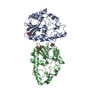
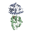
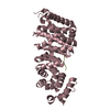
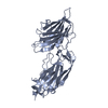
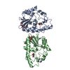
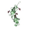
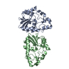
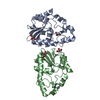
- PDB-1ozn: 1.5A Crystal Structure of the Nogo Receptor Ligand Binding Domain... -
+ Open data

- Basic information
| Entry | Database: PDB / ID: 1ozn | |||||||||
|---|---|---|---|---|---|---|---|---|---|---|
| Title | 1.5A Crystal Structure of the Nogo Receptor Ligand Binding Domain Reveals a Convergent Recognition Scaffold Mediating Inhibition of Myelination | |||||||||
 Components Components | Reticulon 4 receptor | |||||||||
 Keywords Keywords | SIGNALING PROTEIN / Nogo Receptor / MAD / Myelination Inhibition / Omgp / MAG / nogo-66 / p75 / Signal Transduction / Neuronal Regeneration / Ligand Binding | |||||||||
| Function / homology |  Function and homology information Function and homology informationneuronal signal transduction / ganglioside GM1 binding / chondroitin sulfate binding / neuregulin receptor activity / ganglioside GT1b binding / negative regulation of axon regeneration / negative regulation of axon extension / Axonal growth inhibition (RHOA activation) / corpus callosum development / positive regulation of Rho protein signal transduction ...neuronal signal transduction / ganglioside GM1 binding / chondroitin sulfate binding / neuregulin receptor activity / ganglioside GT1b binding / negative regulation of axon regeneration / negative regulation of axon extension / Axonal growth inhibition (RHOA activation) / corpus callosum development / positive regulation of Rho protein signal transduction / axonal growth cone / axonogenesis / dendritic shaft / positive regulation of GTPase activity / negative regulation of neuron projection development / presynapse / signaling receptor activity / heparin binding / perikaryon / cell surface receptor signaling pathway / neuron projection / membrane raft / external side of plasma membrane / neuronal cell body / glutamatergic synapse / protein-containing complex binding / cell surface / endoplasmic reticulum / extracellular exosome / plasma membraneSimilarity search - Function | |||||||||
| Biological species |  Homo sapiens (human) Homo sapiens (human) | |||||||||
| Method | X-RAY DIFFRACTION / SYNCHROTRON / MAD / Resolution: 1.52 Å | |||||||||
 Authors Authors | He, X. / Bazan, J.F. / Park, J.B. / McDermott, G. / He, Z. / Garcia, K.C. | |||||||||
 Citation Citation | Journal: Neuron / Year: 2003 Title: Structure of the Nogo Receptor Ectodomain. A Recognition module implicated in Myelin Inhibition. Authors: He, X.L. / Bazan, J.F. / McDermott, G. / Park, J.B. / Wang, K. / Tessier-Lavigne, M. / He, Z. / Garcia, K.C. | |||||||||
| History |
|
- Structure visualization
| Structure viewer | Molecule: MolmilJmol/JSmol |
|---|
- Downloads & links
-Download
| PDBx/mmCIF format | 1ozn.cif.gz | 76.4 KB | Display | PDBx/mmCIF format |
|---|---|---|---|---|
| PDB format | pdb1ozn.ent.gz | 58.4 KB | Display | PDB format |
| PDBx/mmJSON format | 1ozn.json.gz | Tree view | PDBx/mmJSON format | |
| Others |  Other downloads Other downloads |
-Validation report
| Arichive directory | https://data.pdbj.org/pub/pdb/validation_reports/oz/1oznftp://data.pdbj.org/pub/pdb/validation_reports/oz/1ozn | HTTPS FTP |
|---|
-Related structure data
| Similar structure data |
|---|


-Links
 PDBj
PDBj


- Assembly
| Deposited unit | 
| ||||||||
|---|---|---|---|---|---|---|---|---|---|
| 1 |
| ||||||||
| Unit cell |
| ||||||||
| Details | The Nogo Receptor ligand binding domain binds myelin inhibtors such as Nogo, Omgp and MAG |
-Components
| #1: Protein | / Nogo receptor / NgR / Nogo-66 receptor Mass: 31613.375 Da / Num. of mol.: 1 / Fragment: Ligand Binding Domain Source method: isolated from a genetically manipulated source Source: (gene. exp.) Homo sapiens (human) / Gene: RTN4R OR NOGOR / Plasmid: pAcGP67A / Production host:  Trichoplusia ni (cabbage looper) / Strain (production host): Hi5 / References: UniProt: Q9BZR6 Trichoplusia ni (cabbage looper) / Strain (production host): Hi5 / References: UniProt: Q9BZR6 | ||
|---|---|---|---|
| #2: Polysaccharide | alpha-D-mannopyranose-(1-6)-alpha-D-mannopyranose-(1-4)-2-acetamido-2-deoxy-alpha-D-glucopyranose- ...alpha-D-mannopyranose-(1-6)-alpha-D-mannopyranose-(1-4)-2-acetamido-2-deoxy-alpha-D-glucopyranose-(1-4)-2-acetamido-2-deoxy-beta-D-glucopyranose / Mass: 748.682 Da / Num. of mol.: 1 Source method: isolated from a genetically manipulated source | ||
| #3: Polysaccharide | beta-D-mannopyranose-(1-4)-2-acetamido-2-deoxy-alpha-D-glucopyranose-(1-4)-2-acetamido-2-deoxy-beta- ...beta-D-mannopyranose-(1-4)-2-acetamido-2-deoxy-alpha-D-glucopyranose-(1-4)-2-acetamido-2-deoxy-beta-D-glucopyranose / Mass: 586.542 Da / Num. of mol.: 1 Source method: isolated from a genetically manipulated source | ||
| #4: Chemical | Acetic acid  Mass: 60.052 Da / Num. of mol.: 3 / Source method: obtained synthetically / Formula: C2H4O2 Mass: 60.052 Da / Num. of mol.: 3 / Source method: obtained synthetically / Formula: C2H4O2#5: Water | ChemComp-HOH / | Water Mass: 18.015 Da / Num. of mol.: 354 / Source method: isolated from a natural source / Formula: H2O Mass: 18.015 Da / Num. of mol.: 354 / Source method: isolated from a natural source / Formula: H2O |
-Experimental details
-Experiment
| Experiment | Method: X-RAY DIFFRACTION / Number of used crystals: 1 |
|---|
- Sample preparation
| Crystal | Density Matthews: 1.87 Å3/Da / Density % sol: 34.21 % | |||||||||||||||||||||||||||||||||||
|---|---|---|---|---|---|---|---|---|---|---|---|---|---|---|---|---|---|---|---|---|---|---|---|---|---|---|---|---|---|---|---|---|---|---|---|---|
| Crystal grow | Temperature: 298 K / Method: vapor diffusion, sitting drop / pH: 6 Details: PEG4000, sodium chloride, sodium acetate, pH 6, VAPOR DIFFUSION, SITTING DROP, temperature 298K | |||||||||||||||||||||||||||||||||||
| Crystal grow | *PLUS pH: 6.5 | |||||||||||||||||||||||||||||||||||
| Components of the solutions | *PLUS
|
-Data collection
| Diffraction | Mean temperature: 100 K | ||||||||||||
|---|---|---|---|---|---|---|---|---|---|---|---|---|---|
| Diffraction source | Source: SYNCHROTRON / Site: ALS  / Beamline: 8.2.1 / Wavelength: 1.0064, 1.0096, 0.9950 / Beamline: 8.2.1 / Wavelength: 1.0064, 1.0096, 0.9950 | ||||||||||||
| Detector | Type: ADSC QUANTUM 4 / Detector: CCD / Date: Dec 20, 2002 | ||||||||||||
| Radiation | Protocol: MAD / Monochromatic (M) / Laue (L): M / Scattering type: x-ray | ||||||||||||
| Radiation wavelength |
| ||||||||||||
| Reflection | Resolution: 1.52→50 Å / Num. all: 33484 / Num. obs: 33484 / % possible obs: 96.6 % / Observed criterion σ(F): 0 / Observed criterion σ(I): 0 / Redundancy: 14.4 % / Biso Wilson estimate: 16 Å2 / Rmerge(I) obs: 0.053 / Net I/σ(I): 34 | ||||||||||||
| Reflection shell | Resolution: 1.52→1.56 Å / Redundancy: 13.8 % / Rmerge(I) obs: 0.214 / Mean I/σ(I) obs: 7.4 / % possible all: 96.4 | ||||||||||||
| Reflection | *PLUS Lowest resolution: 50 Å | ||||||||||||
| Reflection shell | *PLUS % possible obs: 96.4 % |
- Processing
| Software |
| ||||||||||||||||||||
|---|---|---|---|---|---|---|---|---|---|---|---|---|---|---|---|---|---|---|---|---|---|
| Refinement | Method to determine structure: MAD / Resolution: 1.52→50 Å / Isotropic thermal model: Isotropic / Cross valid method: THROUGHOUT / σ(F): 0 / σ(I): 0 / Stereochemistry target values: Engh & Huber
| ||||||||||||||||||||
| Displacement parameters | Biso mean: 19.5 Å2
| ||||||||||||||||||||
| Refine analyze |
| ||||||||||||||||||||
| Refinement step | Cycle: LAST / Resolution: 1.52→50 Å
| ||||||||||||||||||||
| Refine LS restraints |
| ||||||||||||||||||||
| LS refinement shell | Resolution: 1.52→1.62 Å / Rfactor Rfree error: 0.017
| ||||||||||||||||||||
| Refinement | *PLUS Lowest resolution: 50 Å | ||||||||||||||||||||
| Solvent computation | *PLUS | ||||||||||||||||||||
| Displacement parameters | *PLUS | ||||||||||||||||||||
| Refine LS restraints | *PLUS
| ||||||||||||||||||||
| LS refinement shell | *PLUS Lowest resolution: 1.56 Å / Rfactor Rfree: 0.278 / Rfactor Rwork: 0.226 |
