[English] 日本語
Yorodumi
- PDB-1ot7: Structural Basis for 3-deoxy-CDCA Binding and Activation of FXR -

+
Open data


ID or keywords:

Loading...

-
Basic information

Entry
Database: PDB / ID: 1ot7
TitleStructural Basis for 3-deoxy-CDCA Binding and Activation of FXR
Components
  • Bile Acid Receptor
  • dodecamer peptide fragment of RPGR-interacting protein 1
KeywordsHORMONE/GROWTH FACTOR RECEPTOR / BILE ACID / NUCLEAR RECEPTOR / COACTIVATOR / LIGAND BINDING DOMAIN / FXR / HORMONE-GROWTH FACTOR RECEPTOR COMPLEX
Function / homology
Function and homology information


response to norepinephrine / Endogenous sterols / Synthesis of bile acids and bile salts / Recycling of bile acids and salts / Synthesis of bile acids and bile salts via 27-hydroxycholesterol / Synthesis of bile acids and bile salts via 7alpha-hydroxycholesterol / negative regulation of triglyceride biosynthetic process / regulation of carbohydrate metabolic process / regulation of lipid storage / hepatocyte proliferation ...response to norepinephrine / Endogenous sterols / Synthesis of bile acids and bile salts / Recycling of bile acids and salts / Synthesis of bile acids and bile salts via 27-hydroxycholesterol / Synthesis of bile acids and bile salts via 7alpha-hydroxycholesterol / negative regulation of triglyceride biosynthetic process / regulation of carbohydrate metabolic process / regulation of lipid storage / hepatocyte proliferation / regulation of bile acid secretion / regulation of urea metabolic process / intracellular bile acid receptor signaling pathway / chenodeoxycholic acid binding / positive regulation of phosphatidic acid biosynthetic process / positive regulation of glutamate metabolic process / positive regulation of ammonia assimilation cycle / negative regulation of collagen biosynthetic process / bile acid receptor activity / regulation of low-density lipoprotein particle clearance / intracellular triglyceride homeostasis / cellular response to bile acid / SUMOylation of intracellular receptors / Nuclear Receptor transcription pathway / negative regulation of very-low-density lipoprotein particle remodeling / negative regulation of interleukin-1 production / leukocyte migration involved in inflammatory response / regulation of insulin secretion involved in cellular response to glucose stimulus / cellular response to organonitrogen compound / toll-like receptor 9 signaling pathway / triglyceride homeostasis / negative regulation of monocyte chemotactic protein-1 production / intracellular receptor signaling pathway / bile acid metabolic process / digestive tract development / bile acid and bile salt transport / response to cholesterol / cell-cell junction assembly / bile acid binding / bile acid signaling pathway / negative regulation of interleukin-2 production / cellular response to fatty acid / positive regulation of interleukin-17 production / positive regulation of insulin secretion involved in cellular response to glucose stimulus / intracellular glucose homeostasis / negative regulation of interleukin-6 production / negative regulation of type II interferon production / negative regulation of tumor necrosis factor production / negative regulation of tumor necrosis factor-mediated signaling pathway / fatty acid homeostasis / response to glucose / nuclear retinoid X receptor binding / negative regulation of canonical NF-kappaB signal transduction / positive regulation of insulin receptor signaling pathway / Notch signaling pathway / positive regulation of adipose tissue development / response to nutrient levels / cholesterol homeostasis / liver regeneration / transcription coregulator binding / RNA polymerase II transcription regulatory region sequence-specific DNA binding / peptide binding / euchromatin / response to organic cyclic compound / negative regulation of inflammatory response / response to estrogen / nuclear receptor activity / cellular response to xenobiotic stimulus / glucose homeostasis / DNA-binding transcription activator activity, RNA polymerase II-specific / response to ethanol / cellular response to lipopolysaccharide / sequence-specific DNA binding / response to lipopolysaccharide / transcription by RNA polymerase II / cell differentiation / receptor complex / DNA-binding transcription factor activity, RNA polymerase II-specific / defense response to bacterium / response to xenobiotic stimulus / RNA polymerase II cis-regulatory region sequence-specific DNA binding / DNA-binding transcription factor activity / innate immune response / protein-containing complex binding / positive regulation of gene expression / regulation of transcription by RNA polymerase II / negative regulation of apoptotic process / positive regulation of DNA-templated transcription / negative regulation of transcription by RNA polymerase II / positive regulation of transcription by RNA polymerase II / DNA binding / zinc ion binding / nucleus
Similarity search - Function
Bile acid receptor, ligand binding domain / Thyroid hormone receptor / Retinoid X Receptor / Retinoid X Receptor / Nuclear hormone receptor / Nuclear hormones receptors DNA-binding region signature. / Zinc finger, nuclear hormone receptor-type / Zinc finger, C4 type (two domains) / Nuclear hormone receptors DNA-binding domain profile. / c4 zinc finger in nuclear hormone receptors ...Bile acid receptor, ligand binding domain / Thyroid hormone receptor / Retinoid X Receptor / Retinoid X Receptor / Nuclear hormone receptor / Nuclear hormones receptors DNA-binding region signature. / Zinc finger, nuclear hormone receptor-type / Zinc finger, C4 type (two domains) / Nuclear hormone receptors DNA-binding domain profile. / c4 zinc finger in nuclear hormone receptors / Nuclear hormone receptor, ligand-binding domain / Nuclear hormone receptor-like domain superfamily / Ligand-binding domain of nuclear hormone receptor / Nuclear receptor (NR) ligand-binding (LBD) domain profile. / Ligand binding domain of hormone receptors / Zinc finger, NHR/GATA-type / Orthogonal Bundle / Mainly Alpha
Similarity search - Domain/homology
6-ETHYL-CHENODEOXYCHOLIC ACID / ISO-URSODEOXYCHOLIC ACID / Bile acid receptor
Similarity search - Component
Biological speciesRattus norvegicus (Norway rat)
MethodX-RAY DIFFRACTION / SYNCHROTRON / MOLECULAR REPLACEMENT / Resolution: 2.9 Å
AuthorsMi, L.Z. / Devarakonda, S. / Harp, J.M. / Han, Q. / Pellicciari, R. / Willson, T.M. / Khorasanizadeh, S. / Rastinejad, F.
CitationJournal: Mol.Cell / Year: 2003
Title: Structural Basis for Bile Acid Binding and Activation of the Nuclear Receptor FXR
Authors: Mi, L.Z. / Devarakonda, S. / Harp, J.M. / Han, Q. / Pellicciari, R. / Willson, T.M. / Khorasanizadeh, S. / Rastinejad, F.
History
DepositionMar 21, 2003Deposition site: RCSB / Processing site: RCSB
Revision 1.0Mar 23, 2004Provider: repository / Type: Initial release
Revision 1.1Apr 2, 2008Group: Version format compliance
Revision 1.2Jul 13, 2011Group: Version format compliance
Revision 1.3Feb 14, 2024Group: Data collection / Database references / Derived calculations
Category: chem_comp_atom / chem_comp_bond ...chem_comp_atom / chem_comp_bond / database_2 / struct_site
Item: _database_2.pdbx_DOI / _database_2.pdbx_database_accession ..._database_2.pdbx_DOI / _database_2.pdbx_database_accession / _struct_site.pdbx_auth_asym_id / _struct_site.pdbx_auth_comp_id / _struct_site.pdbx_auth_seq_id
Revision 1.4Apr 3, 2024Group: Refinement description / Category: pdbx_initial_refinement_model

-
Structure visualization

Structure viewerMolecule:
MolmilJmol/JSmol

Downloads & links

-
Assembly

Deposited unit
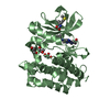
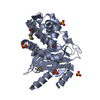
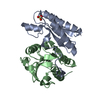
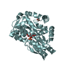
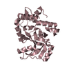
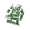
A: Bile Acid Receptor
B: Bile Acid Receptor
C: dodecamer peptide fragment of RPGR-interacting protein 1
D: dodecamer peptide fragment of RPGR-interacting protein 1
E: dodecamer peptide fragment of RPGR-interacting protein 1
hetero molecules


Theoretical massNumber of molelcules
Total (without water)58,7137
Polymers57,9005
Non-polymers8132
Water50428
1
A: Bile Acid Receptor
C: dodecamer peptide fragment of RPGR-interacting protein 1
hetero molecules


Theoretical massNumber of molelcules
Total (without water)28,6383
Polymers28,2172
Non-polymers4211
Water181
TypeNameSymmetry operationNumber
identity operation1_555x,y,z1
2
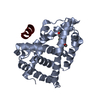
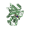
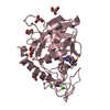
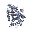
B: Bile Acid Receptor
D: dodecamer peptide fragment of RPGR-interacting protein 1
E: dodecamer peptide fragment of RPGR-interacting protein 1
hetero molecules


Theoretical massNumber of molelcules
Total (without water)30,0754
Polymers29,6823
Non-polymers3931
Water362
TypeNameSymmetry operationNumber
identity operation1_555x,y,z1
Unit cell
Length a, b, c (Å)99.536, 107.129, 69.203
Angle α, β, γ (deg.)90.00, 90.00, 90.00
Int Tables number18
Cell settingorthorhombic
Space group name H-MP21212
DetailsThe biological assembly is one of the subunits(A or B).

-
Components

#1: Protein Bile Acid Receptor / FXR / Farnesoid X-activated receptor / Farnesol receptor HRR-1 / Retinoid X receptor-interacting ...FXR / Farnesoid X-activated receptor / Farnesol receptor HRR-1 / Retinoid X receptor-interacting protein 14 / RXR-interacting protein 14


Mass: 26752.787 Da / Num. of mol.: 2 / Fragment: ligand binding domain
Source method: isolated from a genetically manipulated source
Source: (gene. exp.) Rattus norvegicus (Norway rat) / Gene: NR1H4 / Plasmid: pET-16b / Species (production host): Escherichia coli / Production host: Escherichia coli BL21(DE3) (bacteria) / Strain (production host): BL21(DE3) / References: UniProt: Q62735
#2: Protein/peptide dodecamer peptide fragment of RPGR-interacting protein 1


Mass: 1464.664 Da / Num. of mol.: 3 / Fragment: GRIP1 NID3 / Source method: obtained synthetically / Details: This sequence occurs naturally in humans
#3: Chemical ChemComp-CHC / 6-ETHYL-CHENODEOXYCHOLIC ACID / Obeticholic acid


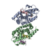
Mass: 420.625 Da / Num. of mol.: 1 / Source method: obtained synthetically / Formula: C26H44O4 / Comment: medication*YM
#4: Chemical ChemComp-IU5 / ISO-URSODEOXYCHOLIC ACID


Mass: 392.572 Da / Num. of mol.: 1 / Source method: obtained synthetically / Formula: C24H40O4
#5: Water ChemComp-HOH / water / Water


Mass: 18.015 Da / Num. of mol.: 28 / Source method: isolated from a natural source / Formula: H2O

-
Experimental details

-
Experiment

ExperimentMethod: X-RAY DIFFRACTION / Number of used crystals: 1

-
Sample preparation

CrystalDensity Matthews: 3.18 Å3/Da / Density % sol: 61 %
Crystal growTemperature: 282 K / Method: vapor diffusion, hanging drop / pH: 6.4
Details: PEG 8000, Ethylene glycol, PIPES, Ammonium Sulfate, pH 6.4, VAPOR DIFFUSION, HANGING DROP, temperature 282K
Crystal grow
*PLUS
pH: 8 / Method: vapor diffusion, hanging drop
Components of the solutions
*PLUS
IDConc.Common nameCrystal-IDSol-IDChemical formulaDetails
1400 mM1dropNaCl
210 mMimidazole1drop
310 %glycerol1drop
420 mMTris1droppH8.0
54-5 mg/mlprotein1drop
70.1 MPIPES1reservoirpH6.4
6ethylene gylcol1reservoir

-
Data collection

DiffractionMean temperature: 100 K
Diffraction sourceSource: SYNCHROTRON / Site: NSLS / Beamline: X4A / Wavelength: 1.067 Å
DetectorType: ADSC QUANTUM 4 / Detector: CCD / Date: Oct 25, 2002
RadiationMonochromator: Si 111 CHANNEL / Protocol: SINGLE WAVELENGTH / Monochromatic (M) / Laue (L): M / Scattering type: x-ray
Radiation wavelengthWavelength: 1.067 Å / Relative weight: 1
ReflectionResolution: 2.9→50 Å / Num. all: 17120 / Num. obs: 16726 / % possible obs: 97.7 % / Observed criterion σ(I): 2.5 / Redundancy: 4.7 % / Biso Wilson estimate: 44.5 Å2 / Rsym value: 0.071 / Net I/σ(I): 14.41
Reflection shellResolution: 2.9→3 Å / Redundancy: 4.8 % / Mean I/σ(I) obs: 2 / Num. unique all: 1612 / Rsym value: 0.458 / % possible all: 96.5
Reflection
*PLUS
Lowest resolution: 15 Å / Num. obs: 12668 / % possible obs: 98.9 % / Num. measured all: 99535 / Rmerge(I) obs: 0.071
Reflection shell
*PLUS
Highest resolution: 2.9 Å / Lowest resolution: 3 Å / % possible obs: 99.6 % / Rmerge(I) obs: 0.458

-
Processing

Software
NameVersionClassification
HKL-2000data collection
SCALEPACKdata scaling
MOLREPphasing
CNS1.1refinement
HKL-2000data reduction
RefinementMethod to determine structure: MOLECULAR REPLACEMENT
Starting model: RAR LBD

Resolution: 2.9→19.96 Å / Rfactor Rfree error: 0.009 / Isotropic thermal model: GROUP / Cross valid method: THROUGHOUT / σ(F): 3 / Stereochemistry target values: Engh & Huber
RfactorNum. reflection% reflectionSelection details
Rfree0.279 880 6.9 %RANDOM
Rwork0.233 ---
all0.236 16938 --
obs0.2331 12771 75.4 %-
Solvent computationSolvent model: FLAT MODEL / Bsol: 18.1809 Å2 / ksol: 0.244014 e/Å3
Displacement parametersBiso mean: 82.9 Å2
Baniso -1Baniso -2Baniso -3
1--9.9 Å20 Å20 Å2
2--21.86 Å20 Å2
3----11.96 Å2
Refine analyzeLuzzati coordinate error free: 0.55 Å / Luzzati sigma a free: 0.46 Å
Refinement stepCycle: LAST / Resolution: 2.9→19.96 Å
ProteinNucleic acidLigandSolventTotal
Num. atoms4055 0 57 28 4140
Refine LS restraints
Refine-IDTypeDev ideal
X-RAY DIFFRACTIONc_bond_d0.009
X-RAY DIFFRACTIONc_bond_d_na
X-RAY DIFFRACTIONc_bond_d_prot
X-RAY DIFFRACTIONc_angle_d
X-RAY DIFFRACTIONc_angle_d_na
X-RAY DIFFRACTIONc_angle_d_prot
X-RAY DIFFRACTIONc_angle_deg1.5
X-RAY DIFFRACTIONc_angle_deg_na
X-RAY DIFFRACTIONc_angle_deg_prot
X-RAY DIFFRACTIONc_dihedral_angle_d19.7
X-RAY DIFFRACTIONc_dihedral_angle_d_na
X-RAY DIFFRACTIONc_dihedral_angle_d_prot
X-RAY DIFFRACTIONc_improper_angle_d0.95
X-RAY DIFFRACTIONc_improper_angle_d_na
X-RAY DIFFRACTIONc_improper_angle_d_prot
X-RAY DIFFRACTIONc_mcbond_it
X-RAY DIFFRACTIONc_mcangle_it
X-RAY DIFFRACTIONc_scbond_it
X-RAY DIFFRACTIONc_scangle_it
LS refinement shellResolution: 2.9→3.03 Å / Rfactor Rfree error: 0.052 / Total num. of bins used: 8
RfactorNum. reflection% reflection
Rfree0.383 55 6 %
Rwork0.371 862 -
obs-862 43.9 %
Xplor file
Refine-IDSerial noParam fileTopol file
X-RAY DIFFRACTION1PROTEIN_REP.PARAMPROTEIN.TOP
X-RAY DIFFRACTION2WATER_REP.PARAMWATER.TOP
X-RAY DIFFRACTION36ECDCA.PAR6ECDCA.TOP
X-RAY DIFFRACTION47ACDCA.PAR7ACDCA.TOP
Refinement
*PLUS
Lowest resolution: 15 Å / % reflection Rfree: 5 % / Rfactor Rfree: 0.29 / Rfactor Rwork: 0.254
Solvent computation
*PLUS
Displacement parameters
*PLUS
Refine LS restraints
*PLUS
Refine-IDTypeDev ideal
X-RAY DIFFRACTIONc_bond_d0.011
X-RAY DIFFRACTIONc_angle_deg1.9
X-RAY DIFFRACTIONc_dihedral_angle_d
X-RAY DIFFRACTIONc_dihedral_angle_deg22.3
X-RAY DIFFRACTIONc_improper_angle_d
X-RAY DIFFRACTIONc_improper_angle_deg1.1

+
About Yorodumi

-
News

-
Feb 9, 2022. New format data for meta-information of EMDB entries

New format data for meta-information of EMDB entries

  • Version 3 of the EMDB header file is now the official format.
  • The previous official version 1.9 will be removed from the archive.

Related info.:EMDB header

External links:wwPDB to switch to version 3 of the EMDB data model

-
Aug 12, 2020. Covid-19 info

Covid-19 info

URL: https://pdbj.org/emnavi/covid19.php

New page: Covid-19 featured information page in EM Navigator.

Related info.:Covid-19 info / Mar 5, 2020. Novel coronavirus structure data

+
Mar 5, 2020. Novel coronavirus structure data

Novel coronavirus structure data

Related info.:Yorodumi Speices / Aug 12, 2020. Covid-19 info

External links:COVID-19 featured content - PDBj / Molecule of the Month (242):Coronavirus Proteases

+
Jan 31, 2019. EMDB accession codes are about to change! (news from PDBe EMDB page)

EMDB accession codes are about to change! (news from PDBe EMDB page)

  • The allocation of 4 digits for EMDB accession codes will soon come to an end. Whilst these codes will remain in use, new EMDB accession codes will include an additional digit and will expand incrementally as the available range of codes is exhausted. The current 4-digit format prefixed with “EMD-” (i.e. EMD-XXXX) will advance to a 5-digit format (i.e. EMD-XXXXX), and so on. It is currently estimated that the 4-digit codes will be depleted around Spring 2019, at which point the 5-digit format will come into force.
  • The EM Navigator/Yorodumi systems omit the EMD- prefix.

Related info.:Q: What is EMD? / ID/Accession-code notation in Yorodumi/EM Navigator

External links:EMDB Accession Codes are Changing Soon! / Contact to PDBj

+
Jul 12, 2017. Major update of PDB

Major update of PDB

  • wwPDB released updated PDB data conforming to the new PDBx/mmCIF dictionary.
  • This is a major update changing the version number from 4 to 5, and with Remediation, in which all the entries are updated.
  • In this update, many items about electron microscopy experimental information are reorganized (e.g. em_software).
  • Now, EM Navigator and Yorodumi are based on the updated data.

External links:wwPDB Remediation / Enriched Model Files Conforming to OneDep Data Standards Now Available in the PDB FTP Archive

-
Yorodumi

Thousand views of thousand structures

  • Yorodumi is a browser for structure data from EMDB, PDB, SASBDB, etc.
  • This page is also the successor to EM Navigator detail page, and also detail information page/front-end page for Omokage search.
  • The word "yorodu" (or yorozu) is an old Japanese word meaning "ten thousand". "mi" (miru) is to see.

Related info.:EMDB / PDB / SASBDB / Comparison of 3 databanks / Yorodumi Search / Aug 31, 2016. New EM Navigator & Yorodumi / Yorodumi Papers / Jmol/JSmol / Function and homology information / Changes in new EM Navigator and Yorodumi

Read more