 Movie
Movie Controller

[English] 日本語
 Yorodumi
- PDB-1o4w: CRYSTAL STRUCTURE OF a PIN (PILT N-TERMINUS) DOMAIN CONTAINING PR... -
+ Open data

- Basic information
| Entry | Database: PDB / ID: 1o4w | ||||||
|---|---|---|---|---|---|---|---|
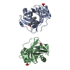
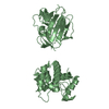
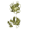
| Title | CRYSTAL STRUCTURE OF a PIN (PILT N-TERMINUS) DOMAIN CONTAINING PROTEIN (AF0591) FROM ARCHAEOGLOBUS FULGIDUS AT 1.90 A RESOLUTION | ||||||
 Components Components | PIN (PilT N-terminus) domain | ||||||
 Keywords Keywords |  TRANSLATION / PIN (PILT N-TERMINUS) DOMAIN / STRUCTURAL GENOMICS / JOINT CENTER FOR STRUCTURAL GENOMICS / JCSG / PROTEIN STRUCTURE INITIATIVE / PSI TRANSLATION / PIN (PILT N-TERMINUS) DOMAIN / STRUCTURAL GENOMICS / JOINT CENTER FOR STRUCTURAL GENOMICS / JCSG / PROTEIN STRUCTURE INITIATIVE / PSI | ||||||
| Function / homology |  Function and homology information Function and homology informationRNA nuclease activity / Hydrolases; Acting on ester bonds / magnesium ion bindingSimilarity search - Function | ||||||
| Biological species |   Archaeoglobus fulgidus (archaea) Archaeoglobus fulgidus (archaea) | ||||||
| Method | X-RAY DIFFRACTION / SYNCHROTRON / MAD / Resolution: 1.9 Å | ||||||
 Authors Authors | Joint Center for Structural Genomics (JCSG) | ||||||
 Citation Citation | Journal: Proteins / Year: 2004 Title: Crystal structure of a PIN (PilT N-terminus) domain (AF0591) from Archaeoglobus fulgidus at 1.90 A resolution Authors: Levin, I. / Schwarzenbacher, R. / Page, R. / Abdubek, P. / Ambing, E. / Biorac, T. / Brinen, L.S. / Campbell, J. / Canaves, J.M. / Chiu, H.J. / Dai, X. / Deacon, A.M. / DiDonato, M. / ...Authors: Levin, I. / Schwarzenbacher, R. / Page, R. / Abdubek, P. / Ambing, E. / Biorac, T. / Brinen, L.S. / Campbell, J. / Canaves, J.M. / Chiu, H.J. / Dai, X. / Deacon, A.M. / DiDonato, M. / Elsliger, M.A. / Floyd, R. / Godzik, A. / Grittini, C. / Grzechnik, S.K. / Hampton, E. / Jaroszewski, L. / Karlak, C. / Klock, H.E. / Koesema, E. / Kovarik, J.S. / Kreusch, A. / Kuhn, P. / Lesley, S.A. / McMullan, D. / McPhillips, T.M. / Miller, M.D. / Morse, A. / Moy, K. / Ouyang, J. / Quijano, K. / Reyes, R. / Rezezadeh, F. / Robb, A. / Sims, E. / Spraggon, G. / Stevens, R.C. / van den Bedem, H. / Velasquez, J. / Vincent, J. / von Delft, F. / Wang, X. / West, B. / Wolf, G. / Xu, Q. / Hodgson, K.O. / Wooley, J. / Wilson, I.A. | ||||||
| History |
| ||||||
| Remark 300 | BIOMOLECULE: 1 THIS ENTRY CONTAINS THE CRYSTALLOGRAPHIC ASYMMETRIC UNIT WHICH CONSISTS OF 1 CHAIN(S) ...BIOMOLECULE: 1 THIS ENTRY CONTAINS THE CRYSTALLOGRAPHIC ASYMMETRIC UNIT WHICH CONSISTS OF 1 CHAIN(S). SEE REMARK 350 FOR INFORMATION ON GENERATING THE BIOLOGICAL MOLECULE(S). THE BIOLOGICAL UNIT IS PROPOSED TO BE A DIMER AROUND THE CRYTALLOGRAPHIC 2-FOLD AXIS. DIMERISATION IS MEDIATED BY AN EXPOSED 2-STRAND BETA SHEET COMPRISING THE HYDROPHILIC C-TERMINAL RESIDUES OF EACH SUBUNIT, THAT BRIDGES THE GLOBULAR DOMAINS OF EACH SUBUNIT. THIS IS CONSIDERED BIOLOGICALLY SIGNIFICANT BECAUSE OF IT LEADS TO CHAIN SWAPPING BETWEEN SUBUNITS. | ||||||
| Remark 650 | HELIX DETERMINATION METHOD: AUTHOR | ||||||
| Remark 700 | SHEET DETERMINATION METHOD: AUTHOR |
- Structure visualization
| Structure viewer | Molecule: MolmilJmol/JSmol |
|---|
- Downloads & links
-Download
| PDBx/mmCIF format | 1o4w.cif.gz | 38.3 KB | Display | PDBx/mmCIF format |
|---|---|---|---|---|
| PDB format | pdb1o4w.ent.gz | 28.1 KB | Display | PDB format |
| PDBx/mmJSON format | 1o4w.json.gz | Tree view | PDBx/mmJSON format | |
| Others |  Other downloads Other downloads |
-Validation report
| Arichive directory | https://data.pdbj.org/pub/pdb/validation_reports/o4/1o4wftp://data.pdbj.org/pub/pdb/validation_reports/o4/1o4w | HTTPS FTP |
|---|
-Related structure data
| Similar structure data | |
|---|---|
| Other databases |


-Links
 PDBj
PDBj

- Assembly
| Deposited unit | 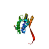
| ||||||||
|---|---|---|---|---|---|---|---|---|---|
| 1 | 
| ||||||||
| Unit cell |
| ||||||||
| Details | THE BIOLOGICAL UNIT IS PROPOSED TO BE A DIMER AROUND THE CRYTALLOGRAPHIC 2-FOLD AXIS. DIMERISATION IS MEDIATED BY AN EXPOSED 2-STRAND BETA SHEET COMPRISING THE HYDROPHILIC C-TERMINAL RESIDUES OF EACH SUBUNIT, THAT BRIDGES THE GLOBULAR DOMAINS OF EACH SUBUNIT. THIS IS CONSIDERED BIOLOGICALLY SIGNIFICANT BECAUSE OF IT LEADS TO CHAIN SWAPPING BETWEEN SUBUNITS. GENERATING THE BIOMOLECULE COORDINATES FOR A COMPLETE MULTIMER REPRESENTING THE KNOWN BIOLOGICALLY SIGNIFICANT OLIGOMERIZATION STATE OF THE MOLECULE CAN BE GENERATED BY APPLYING BIOMT TRANSFORMATIONS GIVEN BELOW. BOTH NON-CRYSTALLOGRAPHIC AND CRYSTALLOGRAPHIC OPERATIONS ARE GIVEN. APPLY THE FOLLOWING TO CHAINS: A, W BIOMT1 1 1.000000 0.000000 0.000000 0.00000 BIOMT2 1 0.000000 1.000000 0.000000 0.00000 BIOMT3 1 0.000000 0.000000 1.000000 0.00000 BIOMT1 2 1.000000 0.000000 0.000000 0.00000 BIOMT2 2 0.000000 -1.000000 0.000000 110.69801 BIOMT3 2 0.000000 0.000000 -1.000000 -26.13300 |
-Components
| #1: Protein | Mass: 17142.221 Da / Num. of mol.: 1 Source method: isolated from a genetically manipulated source Source: (gene. exp.) Archaeoglobus fulgidus (archaea) / Gene: AF0591 / Production host:  Escherichia coli (E. coli) / References: UniProt: O29664 Escherichia coli (E. coli) / References: UniProt: O29664 |
|---|---|
| #2: Water | ChemComp-HOH / Water Mass: 18.015 Da / Num. of mol.: 90 / Source method: isolated from a natural source / Formula: H2O Mass: 18.015 Da / Num. of mol.: 90 / Source method: isolated from a natural source / Formula: H2O |
-Experimental details
-Experiment
| Experiment | Method: X-RAY DIFFRACTION / Number of used crystals: 1 |
|---|
- Sample preparation
| Crystal | Density Matthews: 3.04 Å3/Da / Density % sol: 59.23 % |
|---|---|
| Crystal grow | Temperature: 277 K / Method: vapor diffusion, sitting drop, nanodrop / pH: 6 Details: 30% MPD, MES buffer pH 6.0, VAPOR DIFFUSION,SITTING DROP,NANODROP, temperature 277K |
| Crystal grow | *PLUS Details: Santarsiero, B.D., (2002) J. Appl. Crystallogr., 35, 278. |
-Data collection
| Diffraction | Mean temperature: 100 K | |||||||||||||||
|---|---|---|---|---|---|---|---|---|---|---|---|---|---|---|---|---|
| Diffraction source | Source: SYNCHROTRON / Site: SSRL  / Beamline: BL11-1 / Wavelength: 0.98397, 0.97922, 0.91837, 0.97855 / Beamline: BL11-1 / Wavelength: 0.98397, 0.97922, 0.91837, 0.97855 | |||||||||||||||
| Detector | Type: ADSC QUANTUM 315 / Detector: CCD / Date: Apr 17, 2003 / Details: flat mirror | |||||||||||||||
| Radiation | Monochromator: single crystal Si(111) bent monochromator / Protocol: MAD / Monochromatic (M) / Laue (L): M / Scattering type: x-ray | |||||||||||||||
| Radiation wavelength |
| |||||||||||||||
| Reflection | Resolution: 1.9→31.86 Å / Num. all: 14860 / Num. obs: 14860 / % possible obs: 99.9 % / Redundancy: 8.8 % / Biso Wilson estimate: 48.07 Å2 / Rsym value: 0.05 / Net I/σ(I): 25.3 | |||||||||||||||
| Reflection shell | Resolution: 1.9→1.95 Å / Redundancy: 4.9 % / Mean I/σ(I) obs: 2.3 / Num. unique all: 1058 / Rsym value: 0.638 / % possible all: 99.9 | |||||||||||||||
| Reflection | *PLUS Highest resolution: 1.9 Å / Num. obs: 14887 / Num. measured all: 131380 / Rmerge(I) obs: 0.05 | |||||||||||||||
| Reflection shell | *PLUS % possible obs: 99.9 % / Rmerge(I) obs: 0.638 |
- Processing
| Software |
| |||||||||||||||||||||||||||||||||||||||||||||||||||||||||||||||||||||||||||||||||||||||||||||||||||||||||||||||||||
|---|---|---|---|---|---|---|---|---|---|---|---|---|---|---|---|---|---|---|---|---|---|---|---|---|---|---|---|---|---|---|---|---|---|---|---|---|---|---|---|---|---|---|---|---|---|---|---|---|---|---|---|---|---|---|---|---|---|---|---|---|---|---|---|---|---|---|---|---|---|---|---|---|---|---|---|---|---|---|---|---|---|---|---|---|---|---|---|---|---|---|---|---|---|---|---|---|---|---|---|---|---|---|---|---|---|---|---|---|---|---|---|---|---|---|---|---|
| Refinement | Method to determine structure: MAD / Resolution: 1.9→31.86 Å / Cor.coef. Fo:Fc: 0.965 / Cor.coef. Fo:Fc free: 0.932 / SU B: 6.521 / SU ML: 0.094 / TLS residual ADP flag: LIKELY RESIDUAL / Isotropic thermal model: ISOTROPIC / Cross valid method: THROUGHOUT / ESU R: 0.121 / ESU R Free: 0.125 / Stereochemistry target values: MAXIMUM LIKELIHOOD Details: 1. The 9-sigma difference density peak on the crystallographic 2-fold axis between residues 126 and 128 was left unmodeled, but could indicate a metal ion. 2. The 2 TLS groups correspond to ...Details: 1. The 9-sigma difference density peak on the crystallographic 2-fold axis between residues 126 and 128 was left unmodeled, but could indicate a metal ion. 2. The 2 TLS groups correspond to globular and tail portions of protein respectively. 3. Hydrogens have been added in the riding positions.
| |||||||||||||||||||||||||||||||||||||||||||||||||||||||||||||||||||||||||||||||||||||||||||||||||||||||||||||||||||
| Solvent computation | Ion probe radii: 0.8 Å / Shrinkage radii: 0.8 Å / VDW probe radii: 1.4 Å / Solvent model: BABINET MODEL WITH MASK | |||||||||||||||||||||||||||||||||||||||||||||||||||||||||||||||||||||||||||||||||||||||||||||||||||||||||||||||||||
| Displacement parameters | Biso mean: 41.843 Å2
| |||||||||||||||||||||||||||||||||||||||||||||||||||||||||||||||||||||||||||||||||||||||||||||||||||||||||||||||||||
| Refinement step | Cycle: LAST / Resolution: 1.9→31.86 Å
| |||||||||||||||||||||||||||||||||||||||||||||||||||||||||||||||||||||||||||||||||||||||||||||||||||||||||||||||||||
| Refine LS restraints |
| |||||||||||||||||||||||||||||||||||||||||||||||||||||||||||||||||||||||||||||||||||||||||||||||||||||||||||||||||||
| LS refinement shell | Resolution: 1.9→1.949 Å / Total num. of bins used: 20
| |||||||||||||||||||||||||||||||||||||||||||||||||||||||||||||||||||||||||||||||||||||||||||||||||||||||||||||||||||
| Refinement TLS params. | Method: refined / Refine-ID: X-RAY DIFFRACTION
| |||||||||||||||||||||||||||||||||||||||||||||||||||||||||||||||||||||||||||||||||||||||||||||||||||||||||||||||||||
| Refinement TLS group | Refine-ID: X-RAY DIFFRACTION / Selection: ALL / Auth asym-ID: A / Label asym-ID: A
| |||||||||||||||||||||||||||||||||||||||||||||||||||||||||||||||||||||||||||||||||||||||||||||||||||||||||||||||||||
| Refinement | *PLUS Num. reflection obs: 14859 | |||||||||||||||||||||||||||||||||||||||||||||||||||||||||||||||||||||||||||||||||||||||||||||||||||||||||||||||||||
| Solvent computation | *PLUS | |||||||||||||||||||||||||||||||||||||||||||||||||||||||||||||||||||||||||||||||||||||||||||||||||||||||||||||||||||
| Displacement parameters | *PLUS | |||||||||||||||||||||||||||||||||||||||||||||||||||||||||||||||||||||||||||||||||||||||||||||||||||||||||||||||||||
| Refine LS restraints | *PLUS
|
