 Movie
Movie Controller


+ Open data

- Basic information
| Entry | Database: PDB / ID: 3lgo | ||||||
|---|---|---|---|---|---|---|---|
| Title | Structure of Gse1p, member of the GSE/EGO complex | ||||||
 Components Components | Protein SLM4 | ||||||
 Keywords Keywords | PROTEIN BINDING / Roadblock/LC7 / domain swap / Autophagy / Membrane / Transmembrane / Transport / Vacuole | ||||||
| Function / homology |  Function and homology information Function and homology informationRagulator complex / microautophagy / endocytic recycling / fungal-type vacuole membrane / positive regulation of TOR signaling / late endosome / late endosome membrane / protein transport / signal transduction / identical protein binding / cytoplasm Similarity search - Function | ||||||
| Biological species |  | ||||||
| Method |  X-RAY DIFFRACTION / SYNCHROTRON / Resolution: 2.85 Å X-RAY DIFFRACTION / SYNCHROTRON / Resolution: 2.85 Å | ||||||
 Authors Authors | Kogan, K. / Fass, D. | ||||||
 Citation Citation | Journal: J.Mol.Biol. / Year: 2010 Title: Structural conservation of components in the amino acid sensing branch of the TOR pathway in yeast and mammals. Authors: Kogan, K. / Spear, E.D. / Kaiser, C.A. / Fass, D. | ||||||
| History |
|
- Structure visualization
| Structure viewer | Molecule: MolmilJmol/JSmol |
|---|
- Downloads & links
-Download
| PDBx/mmCIF format | 3lgo.cif.gz | 39.5 KB | Display | PDBx/mmCIF format |
|---|---|---|---|---|
| PDB format | pdb3lgo.ent.gz | 27.3 KB | Display | PDB format |
| PDBx/mmJSON format | 3lgo.json.gz | Tree view | PDBx/mmJSON format | |
| Others |  Other downloads Other downloads |
-Validation report
| Arichive directory | https://data.pdbj.org/pub/pdb/validation_reports/lg/3lgoftp://data.pdbj.org/pub/pdb/validation_reports/lg/3lgo | HTTPS FTP |
|---|
-Related structure data
| Similar structure data |
|---|


-Links
 PDBj
PDBj
- Assembly
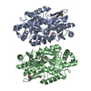
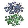
| Deposited unit | 
| ||||||||
|---|---|---|---|---|---|---|---|---|---|
| 1 | 
| ||||||||
| 2 | 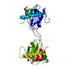
| ||||||||
| Unit cell |
| ||||||||
| Details | AS PER THE AUTHORS THE BIOLOGICAL ASSEMBLY IS UNKNOWN |
-Components
| #1: Protein | Mass: 19563.164 Da / Num. of mol.: 1 Source method: isolated from a genetically manipulated source Source: (gene. exp.) Gene: EGO3, GSE1, SLM4, YBR0723, YBR077C / Plasmid: pet15b / Production host:  |
|---|---|
| #2: Water | ChemComp-HOH /  Mass: 18.015 Da / Num. of mol.: 2 / Source method: isolated from a natural source / Formula: H2O Mass: 18.015 Da / Num. of mol.: 2 / Source method: isolated from a natural source / Formula: H2O |
-Experimental details
-Experiment
| Experiment | Method: X-RAY DIFFRACTION / Number of used crystals: 1 |
|---|
- Sample preparation
| Crystal | Density Matthews: 2.92 Å3/Da / Density % sol: 57.85 % |
|---|---|
| Crystal grow | Temperature: 293 K / Method: vapor diffusion, hanging drop / pH: 3 Details: 100 mM sodium citrate buffer, pH 2.0, 500-800 mM ammonium sulfate, 200 mM lithium sulfate, 200-350 mM L-arginine-HCl, pH 5.75, 5% DMSO, VAPOR DIFFUSION, HANGING DROP, temperature 293K |
-Data collection
| Diffraction | Mean temperature: 100 K |
|---|---|
| Diffraction source | Source: SYNCHROTRON / Site: ESRF  / Beamline: ID14-1 / Wavelength: 0.9334 Å / Beamline: ID14-1 / Wavelength: 0.9334 Å |
| Detector | Type: ADSC QUANTUM 210 / Detector: CCD / Date: Jul 23, 2009 |
| Radiation | Protocol: SINGLE WAVELENGTH / Monochromatic (M) / Laue (L): M / Scattering type: x-ray |
| Radiation wavelength | Wavelength: 0.9334 Å / Relative weight: 1 |
| Reflection | Resolution: 2.85→50 Å / Num. all: 5995 / Num. obs: 5873 / % possible obs: 98.8 % / Redundancy: 13.9 % / Biso Wilson estimate: 83.5 Å2 / Rsym value: 0.065 / Net I/σ(I): 13.8 |
| Reflection shell | Resolution: 2.85→2.9 Å / Redundancy: 14.5 % / Mean I/σ(I) obs: 4 / Rsym value: 0.949 / % possible all: 97.9 |
- Processing
| Software |
| |||||||||||||||||||||||||
|---|---|---|---|---|---|---|---|---|---|---|---|---|---|---|---|---|---|---|---|---|---|---|---|---|---|---|
| Refinement | Resolution: 2.85→50 Å / Cross valid method: THROUGHOUT
| |||||||||||||||||||||||||
| Refinement step | Cycle: LAST / Resolution: 2.85→50 Å
| |||||||||||||||||||||||||
| Refine LS restraints |
|
