 Movie
Movie Controller

[English] 日本語
 Yorodumi
- PDB-1nzi: Crystal Structure of the CUB1-EGF Interaction Domain of Complemen... -
+ Open data

- Basic information
| Entry | Database: PDB / ID: 1nzi | ||||||
|---|---|---|---|---|---|---|---|
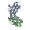
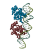
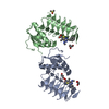
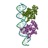
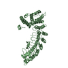
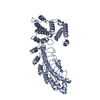
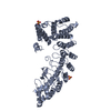
| Title | Crystal Structure of the CUB1-EGF Interaction Domain of Complement Protease C1s | ||||||
 Components Components | Complement C1s component | ||||||
 Keywords Keywords | HYDROLASE / CALCIUM / COMPLEMENT / INNATE IMMUNITY / MODULAR STRUCTURE / CUB / EGF | ||||||
| Function / homology |  Function and homology information Function and homology informationcomplement subcomponent C_overbar_1s_ / Classical antibody-mediated complement activation / Initial triggering of complement / complement activation, classical pathway / Regulation of Complement cascade / blood microparticle / serine-type endopeptidase activity / innate immune response / calcium ion binding / proteolysis ...complement subcomponent C_overbar_1s_ / Classical antibody-mediated complement activation / Initial triggering of complement / complement activation, classical pathway / Regulation of Complement cascade / blood microparticle / serine-type endopeptidase activity / innate immune response / calcium ion binding / proteolysis / extracellular space / extracellular region / identical protein binding Similarity search - Function | ||||||
| Biological species |  Homo sapiens (human) Homo sapiens (human) | ||||||
| Method |  X-RAY DIFFRACTION / SYNCHROTRON / SAD, molecular replacement / Resolution: 1.5 Å X-RAY DIFFRACTION / SYNCHROTRON / SAD, molecular replacement / Resolution: 1.5 Å | ||||||
 Authors Authors | Gregory, L.A. / Thielens, N.M. / Arlaud, G.J. / Fontecilla-Camps, J.C. / Gaboriaud, C. | ||||||
 Citation Citation | Journal: J.Biol.Chem. / Year: 2003 Title: X-ray structure of the Ca2+-binding interaction domain of C1s. Insights into the assembly of the C1 complex of complement Authors: Gregory, L.A. / Thielens, N.M. / Arlaud, G.J. / Fontecilla-Camps, J.C. / Gaboriaud, C. | ||||||
| History |
|
- Structure visualization
| Structure viewer | Molecule: MolmilJmol/JSmol |
|---|
- Downloads & links
-Download
| PDBx/mmCIF format | 1nzi.cif.gz | 78.7 KB | Display | PDBx/mmCIF format |
|---|---|---|---|---|
| PDB format | pdb1nzi.ent.gz | 63 KB | Display | PDB format |
| PDBx/mmJSON format | 1nzi.json.gz | Tree view | PDBx/mmJSON format | |
| Others |  Other downloads Other downloads |
-Validation report
| Summary document | 1nzi_validation.pdf.gz | 438.7 KB | Display | wwPDB validaton report |
|---|---|---|---|---|
| Full document | 1nzi_full_validation.pdf.gz | 441.5 KB | Display | |
| Data in XML | 1nzi_validation.xml.gz | 17 KB | Display | |
| Data in CIF | 1nzi_validation.cif.gz | 24.5 KB | Display | |
| Arichive directory | https://data.pdbj.org/pub/pdb/validation_reports/nz/1nziftp://data.pdbj.org/pub/pdb/validation_reports/nz/1nzi | HTTPS FTP |
-Related structure data
| Similar structure data |
|---|

-Links
 PDBj
PDBj


- Assembly
| Deposited unit | 
| ||||||||
|---|---|---|---|---|---|---|---|---|---|
| 1 |
| ||||||||
| Unit cell |
| ||||||||
| Details | The biological assembly is a calcium-dependent homodimer in the asymmetric unit |
-Components
| #1: Protein | Mass: 18144.895 Da / Num. of mol.: 2 / Fragment: CALCIUM-DEPENDENT INTERACTION DOMAIN Source method: isolated from a genetically manipulated source Source: (gene. exp.) Homo sapiens (human) / Gene: C1S / Cell line (production host): HIGH FIVE INSECT CELLS / Production host:  Trichoplusia ni (cabbage looper) Trichoplusia ni (cabbage looper)References: UniProt: P09871, complement subcomponent C_overbar_1s_ #2: Chemical |   Mass: 40.078 Da / Num. of mol.: 2 / Source method: obtained synthetically / Formula: Ca Mass: 40.078 Da / Num. of mol.: 2 / Source method: obtained synthetically / Formula: Ca#3: Chemical |   Mass: 24.305 Da / Num. of mol.: 2 / Source method: obtained synthetically / Formula: Mg Mass: 24.305 Da / Num. of mol.: 2 / Source method: obtained synthetically / Formula: Mg#4: Water | ChemComp-HOH / |  Mass: 18.015 Da / Num. of mol.: 288 / Source method: isolated from a natural source / Formula: H2O Mass: 18.015 Da / Num. of mol.: 288 / Source method: isolated from a natural source / Formula: H2O |
|---|
-Experimental details
-Experiment
| Experiment | Method: X-RAY DIFFRACTION / Number of used crystals: 1 |
|---|
- Sample preparation
| Crystal | Density Matthews: 2.28 Å3/Da / Density % sol: 45.62 % | ||||||||||||||||||||||||||||||||||||||||||||||||||||||||
|---|---|---|---|---|---|---|---|---|---|---|---|---|---|---|---|---|---|---|---|---|---|---|---|---|---|---|---|---|---|---|---|---|---|---|---|---|---|---|---|---|---|---|---|---|---|---|---|---|---|---|---|---|---|---|---|---|---|
| Crystal grow | Temperature: 293 K / Method: vapor diffusion, hanging drop / pH: 7.5 Details: PEG 4000, magnesium chloride, glycerol, pH 7.5, VAPOR DIFFUSION, HANGING DROP, temperature 293K | ||||||||||||||||||||||||||||||||||||||||||||||||||||||||
| Crystal grow | *PLUS Temperature: 20 ℃ / pH: 7.4 / Method: vapor diffusion, hanging drop | ||||||||||||||||||||||||||||||||||||||||||||||||||||||||
| Components of the solutions | *PLUS
|
-Data collection
| Diffraction | Mean temperature: 100 K |
|---|---|
| Diffraction source | Source: SYNCHROTRON / Site: ESRF  / Beamline: ID14-2 / Wavelength: 0.933 Å / Beamline: ID14-2 / Wavelength: 0.933 Å |
| Detector | Type: MARRESEARCH / Detector: CCD / Date: Apr 21, 2000 |
| Radiation | Protocol: SINGLE WAVELENGTH / Monochromatic (M) / Laue (L): M / Scattering type: x-ray |
| Radiation wavelength | Wavelength: 0.933 Å / Relative weight: 1 |
| Reflection | Resolution: 1.5→30 Å / Num. all: 52134 / Num. obs: 52134 / % possible obs: 94.4 % / Observed criterion σ(F): 0 / Observed criterion σ(I): 0 / Rsym value: 0.064 / Net I/σ(I): 6.9 |
| Reflection shell | Resolution: 1.5→1.58 Å / Rsym value: 0.143 / % possible all: 93.3 |
| Reflection | *PLUS Highest resolution: 1.5 Å / Num. obs: 52518 / Redundancy: 3.5 % / Num. measured all: 181271 / Rmerge(I) obs: 0.064 |
| Reflection shell | *PLUS % possible obs: 93.3 % / Redundancy: 2.2 % / Num. unique obs: 7588 / Num. measured obs: 16763 / Rmerge(I) obs: 0.143 / Mean I/σ(I) obs: 4 |
- Processing
| Software |
| ||||||||||||||||||||
|---|---|---|---|---|---|---|---|---|---|---|---|---|---|---|---|---|---|---|---|---|---|
| Refinement | Method to determine structure: SAD, molecular replacement / Resolution: 1.5→30 Å / Cross valid method: THROUGHOUT / σ(F): 0 / Stereochemistry target values: Engh & Huber
| ||||||||||||||||||||
| Refinement step | Cycle: LAST / Resolution: 1.5→30 Å
| ||||||||||||||||||||
| LS refinement shell | Resolution: 1.5→1.58 Å / Rfactor Rfree error: 0.01
| ||||||||||||||||||||
| Refinement | *PLUS Highest resolution: 1.5 Å / Rfactor Rfree: 0.246 / Rfactor Rwork: 0.229 | ||||||||||||||||||||
| Solvent computation | *PLUS | ||||||||||||||||||||
| Displacement parameters | *PLUS |
