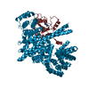
 Movie
Movie Controller


+ Open data

- Basic information
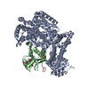
| Entry | Database: PDB / ID: 1n52 | ||||||
|---|---|---|---|---|---|---|---|
| Title | Cap Binding Complex | ||||||
 Components Components |
| ||||||
 Keywords Keywords | RNA BINDING PROTEIN / CBP80 / CBP20 / RNP domain / cap binding protein / m7GpppG | ||||||
| Function / homology |  Function and homology information Function and homology informationpositive regulation of RNA binding / snRNA export from nucleus / nuclear cap binding complex / RNA cap binding complex / histone mRNA metabolic process / mRNA metabolic process / positive regulation of RNA export from nucleus / positive regulation of mRNA 3'-end processing / cap-dependent translational initiation / Processing of Intronless Pre-mRNAs ...positive regulation of RNA binding / snRNA export from nucleus / nuclear cap binding complex / RNA cap binding complex / histone mRNA metabolic process / mRNA metabolic process / positive regulation of RNA export from nucleus / positive regulation of mRNA 3'-end processing / cap-dependent translational initiation / Processing of Intronless Pre-mRNAs / RNA cap binding / snRNA binding / primary miRNA processing / miRNA-mediated post-transcriptional gene silencing / regulation of mRNA processing / SLBP independent Processing of Histone Pre-mRNAs / SLBP Dependent Processing of Replication-Dependent Histone Pre-mRNAs / regulatory ncRNA-mediated post-transcriptional gene silencing / Transport of the SLBP independent Mature mRNA / Transport of the SLBP Dependant Mature mRNA / RNA 7-methylguanosine cap binding / alternative mRNA splicing, via spliceosome / positive regulation of mRNA splicing, via spliceosome / mRNA 3'-end processing / Transport of Mature mRNA Derived from an Intronless Transcript / mRNA 3'-end processing / mRNA cis splicing, via spliceosome / Transport of Mature mRNA derived from an Intron-Containing Transcript / RNA catabolic process / Abortive elongation of HIV-1 transcript in the absence of Tat / RNA Polymerase II Transcription Termination / nuclear-transcribed mRNA catabolic process, nonsense-mediated decay / regulation of translational initiation / FGFR2 alternative splicing / Signaling by FGFR2 IIIa TM / Formation of the Early Elongation Complex / Formation of the HIV-1 Early Elongation Complex / mRNA Capping / spliceosomal complex assembly / mRNA Splicing - Minor Pathway / Processing of Capped Intron-Containing Pre-mRNA / RNA polymerase II transcribes snRNA genes / 7-methylguanosine mRNA capping / Nonsense Mediated Decay (NMD) independent of the Exon Junction Complex (EJC) / mRNA export from nucleus / Formation of HIV-1 elongation complex containing HIV-1 Tat / Formation of HIV elongation complex in the absence of HIV Tat / Nonsense Mediated Decay (NMD) enhanced by the Exon Junction Complex (EJC) / Formation of RNA Pol II elongation complex / RNA Polymerase II Pre-transcription Events / mRNA Splicing - Major Pathway / RNA splicing / positive regulation of transcription elongation by RNA polymerase II / mRNA splicing, via spliceosome / mRNA transcription by RNA polymerase II / Regulation of expression of SLITs and ROBOs / positive regulation of cell growth / snRNP Assembly / defense response to virus / molecular adaptor activity / ciliary basal body / ribonucleoprotein complex / mRNA binding / mitochondrion / DNA binding / RNA binding / nucleoplasm / nucleus / cytoplasm / cytosol Similarity search - Function | ||||||
| Biological species |  Homo sapiens (human) Homo sapiens (human) | ||||||
| Method |  X-RAY DIFFRACTION / SYNCHROTRON / MOLECULAR REPLACEMENT / Resolution: 2.11 Å X-RAY DIFFRACTION / SYNCHROTRON / MOLECULAR REPLACEMENT / Resolution: 2.11 Å | ||||||
 Authors Authors | Calero, G. / Wilson, K. / Ly, T. / Rios-Steiner, J. / Clardy, J. / Cerione, R. | ||||||
 Citation Citation | Journal: Nat.Struct.Biol. / Year: 2002 Title: Structural basis of m7GpppG binding to the nuclear cap-binding protein complex. Authors: Calero, G. / Wilson, K. / Ly, T. / Rios-Steiner, J. / Clardy, J. / Cerione, R. | ||||||
| History |
|
- Structure visualization
| Structure viewer | Molecule: MolmilJmol/JSmol |
|---|
- Downloads & links
-Download
| PDBx/mmCIF format | 1n52.cif.gz | 203.9 KB | Display | PDBx/mmCIF format |
|---|---|---|---|---|
| PDB format | pdb1n52.ent.gz | 159.6 KB | Display | PDB format |
| PDBx/mmJSON format | 1n52.json.gz | Tree view | PDBx/mmJSON format | |
| Others |  Other downloads Other downloads |
-Validation report
| Arichive directory | https://data.pdbj.org/pub/pdb/validation_reports/n5/1n52ftp://data.pdbj.org/pub/pdb/validation_reports/n5/1n52 | HTTPS FTP |
|---|
-Related structure data
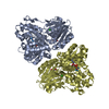
| Related structure data |  1n54C  1h6kS S: Starting model for refinement C: citing same article ( |
|---|---|
| Similar structure data |


-Links
 PDBj
PDBj


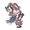
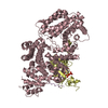
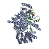
- Assembly
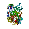
| Deposited unit | 
| ||||||||
|---|---|---|---|---|---|---|---|---|---|
| 1 |
| ||||||||
| Unit cell |
|
-Components
-Protein , 2 types, 2 molecules AB
| #1: Protein | Mass: 91960.297 Da / Num. of mol.: 1 Source method: isolated from a genetically manipulated source Source: (gene. exp.) Homo sapiens (human) / Gene: CBP80 / Cell line (production host): Sf21 / Production host:   Spodoptera frugiperda (fall armyworm) / References: UniProt: Q09161 Spodoptera frugiperda (fall armyworm) / References: UniProt: Q09161 |
|---|---|
| #2: Protein | Mass: 18028.131 Da / Num. of mol.: 1 Source method: isolated from a genetically manipulated source Source: (gene. exp.) Homo sapiens (human) / Gene: CBP20 / Cell line (production host): Sf21 / Production host: Spodoptera frugiperda (fall armyworm) / References: UniProt: P52298 |
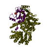
-Non-polymers , 5 types, 357 molecules 


| #3: Chemical | ChemComp-MG /  Mass: 24.305 Da / Num. of mol.: 1 / Source method: obtained synthetically / Formula: Mg Mass: 24.305 Da / Num. of mol.: 1 / Source method: obtained synthetically / Formula: Mg | ||||
|---|---|---|---|---|---|
| #4: Chemical | ChemComp-PG4 /  Mass: 194.226 Da / Num. of mol.: 1 / Source method: obtained synthetically / Formula: C8H18O5 / Comment: precipitant*YM Mass: 194.226 Da / Num. of mol.: 1 / Source method: obtained synthetically / Formula: C8H18O5 / Comment: precipitant*YM | ||||
| #5: Chemical | ChemComp-GOL /  Mass: 92.094 Da / Num. of mol.: 16 / Source method: obtained synthetically / Formula: C3H8O3 Mass: 92.094 Da / Num. of mol.: 16 / Source method: obtained synthetically / Formula: C3H8O3#6: Chemical | ChemComp-GTG / |  Mass: 803.440 Da / Num. of mol.: 1 / Source method: obtained synthetically / Formula: C21H30N10O18P3 Mass: 803.440 Da / Num. of mol.: 1 / Source method: obtained synthetically / Formula: C21H30N10O18P3#7: Water | ChemComp-HOH / | Mass: 18.015 Da / Num. of mol.: 338 / Source method: isolated from a natural source / Formula: H2O |
-Experimental details
-Experiment
| Experiment | Method: X-RAY DIFFRACTION / Number of used crystals: 3 |
|---|
- Sample preparation
| Crystal | Density Matthews: 2.97 Å3/Da / Density % sol: 58.2 % | |||||||||||||||||||||||||||||||||||||||||||||||||||||||||||||||
|---|---|---|---|---|---|---|---|---|---|---|---|---|---|---|---|---|---|---|---|---|---|---|---|---|---|---|---|---|---|---|---|---|---|---|---|---|---|---|---|---|---|---|---|---|---|---|---|---|---|---|---|---|---|---|---|---|---|---|---|---|---|---|---|---|
| Crystal grow | Temperature: 298 K / Method: vapor diffusion, sitting drop / pH: 7.25 Details: PEG 400, magnesium chloride, Tris, Glycine, Ethylene Glycol , pH 7.25, VAPOR DIFFUSION, SITTING DROP, temperature 298K | |||||||||||||||||||||||||||||||||||||||||||||||||||||||||||||||
| Crystal grow | *PLUS Temperature: 4 ℃ | |||||||||||||||||||||||||||||||||||||||||||||||||||||||||||||||
| Components of the solutions | *PLUS
|
-Data collection
| Diffraction |
| |||||||||||||||
|---|---|---|---|---|---|---|---|---|---|---|---|---|---|---|---|---|
| Diffraction source |
| |||||||||||||||
| Detector |
| |||||||||||||||
| Radiation | Protocol: MAD / Monochromatic (M) / Laue (L): M / Scattering type: x-ray | |||||||||||||||
| Radiation wavelength |
| |||||||||||||||
| Reflection | Resolution: 2.12→37.45 Å / Num. all: 135299 / Num. obs: 132887 / % possible obs: 98 % / Observed criterion σ(F): 4 / Observed criterion σ(I): 4 / Redundancy: 6 % / Biso Wilson estimate: 14.9 Å2 / Limit h max: 48 / Limit h min: -27 / Limit k max: 55 / Limit k min: -27 / Limit l max: 88 / Limit l min: -88 / Observed criterion F max: 3903632.29 / Observed criterion F min: 0.2 / Rmerge(I) obs: 0.33 / Rsym value: 0.067 / Net I/σ(I): 16 | |||||||||||||||
| Reflection shell | Resolution: 2.11→2.17 Å / Mean I/σ(I) obs: 2.4 | |||||||||||||||
| Reflection | *PLUS Lowest resolution: 40 Å / Num. obs: 78602 / % possible obs: 99 % / Rmerge(I) obs: 0.058 | |||||||||||||||
| Reflection shell | *PLUS Mean I/σ(I) obs: 2.1 |

- Processing
| Software |
| ||||||||||||||||||||||||||||||||||||||||||||||||||||||||||||||||||||||||||||||||||||||||||
|---|---|---|---|---|---|---|---|---|---|---|---|---|---|---|---|---|---|---|---|---|---|---|---|---|---|---|---|---|---|---|---|---|---|---|---|---|---|---|---|---|---|---|---|---|---|---|---|---|---|---|---|---|---|---|---|---|---|---|---|---|---|---|---|---|---|---|---|---|---|---|---|---|---|---|---|---|---|---|---|---|---|---|---|---|---|---|---|---|---|---|---|
| Refinement | Method to determine structure: MOLECULAR REPLACEMENT Starting model: PDB entry 1H6K Resolution: 2.11→24.54 Å / Rfactor Rfree error: 0.004 / Occupancy max: 1.07 / Occupancy min: 1 / Data cutoff high absF: 3917587.28 / Data cutoff high rms absF: 3917587.28 / Data cutoff low absF: 0 / Cross valid method: THROUGHOUT / σ(F): 4 / Stereochemistry target values: Engh & Huber
| ||||||||||||||||||||||||||||||||||||||||||||||||||||||||||||||||||||||||||||||||||||||||||
| Solvent computation | Solvent model: CNS bulk solvent model used / Bsol: 45.8457 Å2 / ksol: 0.343953 e/Å3 | ||||||||||||||||||||||||||||||||||||||||||||||||||||||||||||||||||||||||||||||||||||||||||
| Displacement parameters | Biso max: 94.13 Å2 / Biso mean: 49.2 Å2 / Biso min: 15.89 Å2
| ||||||||||||||||||||||||||||||||||||||||||||||||||||||||||||||||||||||||||||||||||||||||||
| Refine analyze |
| ||||||||||||||||||||||||||||||||||||||||||||||||||||||||||||||||||||||||||||||||||||||||||
| Refinement step | Cycle: LAST / Resolution: 2.11→24.54 Å
| ||||||||||||||||||||||||||||||||||||||||||||||||||||||||||||||||||||||||||||||||||||||||||
| Refine LS restraints |
| ||||||||||||||||||||||||||||||||||||||||||||||||||||||||||||||||||||||||||||||||||||||||||
| LS refinement shell | Refine-ID: X-RAY DIFFRACTION / Total num. of bins used: 8
| ||||||||||||||||||||||||||||||||||||||||||||||||||||||||||||||||||||||||||||||||||||||||||
| Xplor file |
| ||||||||||||||||||||||||||||||||||||||||||||||||||||||||||||||||||||||||||||||||||||||||||
| Software | *PLUS Name: CNS / Classification: refinement | ||||||||||||||||||||||||||||||||||||||||||||||||||||||||||||||||||||||||||||||||||||||||||
| Refinement | *PLUS Highest resolution: 2.12 Å / Lowest resolution: 40 Å / Rfactor Rfree: 0.2427 / Rfactor Rwork: 0.2256 | ||||||||||||||||||||||||||||||||||||||||||||||||||||||||||||||||||||||||||||||||||||||||||
| Solvent computation | *PLUS | ||||||||||||||||||||||||||||||||||||||||||||||||||||||||||||||||||||||||||||||||||||||||||
| Displacement parameters | *PLUS | ||||||||||||||||||||||||||||||||||||||||||||||||||||||||||||||||||||||||||||||||||||||||||
| Refine LS restraints | *PLUS
| ||||||||||||||||||||||||||||||||||||||||||||||||||||||||||||||||||||||||||||||||||||||||||
| LS refinement shell | *PLUS Rfactor Rfree: 0.37 / Rfactor Rwork: 0.32 |
