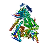
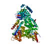
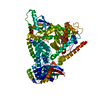
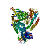
Entry Database : PDB / ID : 5ubrTitle CRYSTAL STRUCTURE OF PI3K ALPHA IN COMPLEX WITH A 7-(3-(PIPERAZIN-1-YL)PHENYL)PYRROLO[2,1-F][1,2,4] TRIAZIN-4-AMINE DERIVIATINE Phosphatidylinositol 4,5-bisphosphate 3-kinase catalytic subunit alpha isoform Keywords / / / / / Function / homology Function Domain/homology Component
/ / / / / / / / / / / / / / / / / / / / / / / / / / / / / / / / / / / / / / / / / / / / / / / / / / / / / / / / / / / / / / / / / / / / / / / / / / / / / / / / / / / / / / / / / / / / / / / / / / / / / / / / / / / / / / / / / / / / / / / / / / / / / / / / / / / / / / / / / / / / / / / / / / / / / / / / / / / / / / Biological species Homo sapiens (human)Method / / / Resolution : 2.4 Å Authors Sack, J.S. Journal : Bioorg. Med. Chem. Lett. / Year : 2017Title : Discovery of 7-(3-(piperazin-1-yl)phenyl)pyrrolo[2,1-f][1,2,4]triazin-4-amine derivatives as highly potent and selective PI3K delta inhibitors.Authors: Qin, L.Y. / Ruan, Z. / Cherney, R.J. / Dhar, T.G. / Neels, J. / Weigelt, C.A. / Sack, J.S. / Srivastava, A.S. / Cornelius, L.A. / Tino, J.A. / Stefanski, K. / Gu, X. / Xie, J. / Susulic, V. ... Authors : Qin, L.Y. / Ruan, Z. / Cherney, R.J. / Dhar, T.G. / Neels, J. / Weigelt, C.A. / Sack, J.S. / Srivastava, A.S. / Cornelius, L.A. / Tino, J.A. / Stefanski, K. / Gu, X. / Xie, J. / Susulic, V. / Yang, X. / Yarde-Chinn, M. / Skala, S. / Bosnius, R. / Goldstein, C. / Davies, P. / Ruepp, S. / Salter-Cid, L. / Bhide, R.S. / Poss, M.A. History Deposition Dec 21, 2016 Deposition site / Processing site Revision 1.0 Feb 8, 2017 Provider / Type Revision 1.1 Feb 22, 2017 Group Revision 1.2 Mar 6, 2024 Group / Database referencesCategory chem_comp_atom / chem_comp_bond ... chem_comp_atom / chem_comp_bond / database_2 / diffrn_radiation_wavelength Item / _database_2.pdbx_database_accession
Show all Show less
 Movie
Movie Controller

 Yorodumi
 Open data
 Basic information
 Components
 Keywords
 Function and homology information
 Homo sapiens (human)
 X-RAY DIFFRACTION /
  Authors
 Citation
 Structure visualization
 Downloads & links
 Other downloads
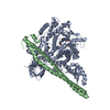


 PDBj
PDBj


 Assembly
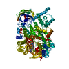
 unidentified baculovirus
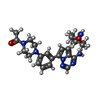
 Mass: 486.569 Da / Num. of mol.: 1 / Source method: obtained synthetically / Formula: C26H30N8O2
 Mass: 18.015 Da / Num. of mol.: 158 / Source method: isolated from a natural source / Formula: H2O
 Sample preparation
 / Beamline: X06SA / Wavelength: 1 Å
 Processing