 Movie
Movie Controller

[English] 日本語
 Yorodumi
- PDB-1mly: Crystal Structure of 7,8-Diaminopelargonic Acid Synthase in compl... -
+ Open data

- Basic information
| Entry | Database: PDB / ID: 1mly | ||||||
|---|---|---|---|---|---|---|---|
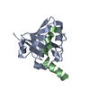
| Title | Crystal Structure of 7,8-Diaminopelargonic Acid Synthase in complex with the cis isomer of amiclenomycin | ||||||
 Components Components | 7,8-diamino-pelargonic acid aminotransferase | ||||||
 Keywords Keywords | TRANSFERASE / Aminotransferase / Fold type I / subclass II / amiclenomycin | ||||||
| Function / homology |  Function and homology information Function and homology informationadenosylmethionine-8-amino-7-oxononanoate transaminase / adenosylmethionine-8-amino-7-oxononanoate transaminase activity / biotin biosynthetic process / pyridoxal phosphate binding / protein homodimerization activity / cytoplasm Similarity search - Function | ||||||
| Biological species |  | ||||||
| Method |  X-RAY DIFFRACTION / SYNCHROTRON / MOLECULAR REPLACEMENT / Resolution: 1.81 Å X-RAY DIFFRACTION / SYNCHROTRON / MOLECULAR REPLACEMENT / Resolution: 1.81 Å | ||||||
 Authors Authors | Sandmark, J. / Mann, S. / Marquet, A. / Schneider, G. | ||||||
 Citation Citation | Journal: J.Biol.Chem. / Year: 2002 Title: Structural basis for the inhibition of the biosynthesis of biotin by the antibiotic amiclenomycin Authors: Sandmark, J. / Mann, S. / Marquet, A. / Schneider, G. | ||||||
| History |
| ||||||
| Remark 999 | sequence The crystallized protein differs from the Swissprot sequence at residue 14. In the ...sequence The crystallized protein differs from the Swissprot sequence at residue 14. In the Swissprot sequence number 14 is a tryptophan while here it is a leucin. This is confirmed by DNA sequencing and was also reported for the original WT structure (1qj5). |
- Structure visualization
| Structure viewer | Molecule: MolmilJmol/JSmol |
|---|
- Downloads & links
-Download
| PDBx/mmCIF format | 1mly.cif.gz | 191.3 KB | Display | PDBx/mmCIF format |
|---|---|---|---|---|
| PDB format | pdb1mly.ent.gz | 150.4 KB | Display | PDB format |
| PDBx/mmJSON format | 1mly.json.gz | Tree view | PDBx/mmJSON format | |
| Others |  Other downloads Other downloads |
-Validation report
| Arichive directory | https://data.pdbj.org/pub/pdb/validation_reports/ml/1mlyftp://data.pdbj.org/pub/pdb/validation_reports/ml/1mly | HTTPS FTP |
|---|
-Related structure data


-Links
 PDBj
PDBj- Assembly
| Deposited unit | 
| ||||||||
|---|---|---|---|---|---|---|---|---|---|
| 1 |
| ||||||||
| Unit cell |
|
-Components
| #1: Protein | Mass: 47310.391 Da / Num. of mol.: 2 Source method: isolated from a genetically manipulated source Source: (gene. exp.) References: UniProt: P12995, adenosylmethionine-8-amino-7-oxononanoate transaminase #2: Chemical |   Mass: 196.246 Da / Num. of mol.: 2 / Source method: obtained synthetically / Formula: C10H16N2O2 Mass: 196.246 Da / Num. of mol.: 2 / Source method: obtained synthetically / Formula: C10H16N2O2#3: Chemical |   Mass: 22.990 Da / Num. of mol.: 2 / Source method: obtained synthetically / Formula: Na Mass: 22.990 Da / Num. of mol.: 2 / Source method: obtained synthetically / Formula: Na#4: Chemical |   Mass: 247.142 Da / Num. of mol.: 2 / Source method: obtained synthetically / Formula: C8H10NO6P Mass: 247.142 Da / Num. of mol.: 2 / Source method: obtained synthetically / Formula: C8H10NO6P#5: Water | ChemComp-HOH / |  Mass: 18.015 Da / Num. of mol.: 537 / Source method: isolated from a natural source / Formula: H2O Mass: 18.015 Da / Num. of mol.: 537 / Source method: isolated from a natural source / Formula: H2ONonpolymer details | In both subunits the cofactor pyridoxal-5'-phosphate forms a covalent adduct with the inhibitor ...In both subunits the cofactor pyridoxal-5'-phosphate forms a covalent adduct with the inhibitor amiclenomycin, followed by aromatization of amiclenomycin hexadiene ring. The binding mode for the inhibitor is different in the two subunits. The electron density for the covalent adduct between amiclenomycin and pyridoxal-5'-phosphate is better in monomer A. For details see the primary citation. | |
|---|
-Experimental details
-Experiment
| Experiment | Method: X-RAY DIFFRACTION / Number of used crystals: 1 |
|---|
- Sample preparation
| Crystal | Density Matthews: 2.08 Å3/Da / Density % sol: 40.91 % | ||||||||||||||||||||
|---|---|---|---|---|---|---|---|---|---|---|---|---|---|---|---|---|---|---|---|---|---|
| Crystal grow | Temperature: 294 K / Method: vapor diffusion, hanging drop / pH: 7.5 Details: PEG4000, MPD, HEPES, pH 7.5, VAPOR DIFFUSION, HANGING DROP, temperature 294K | ||||||||||||||||||||
| Crystal grow | *PLUS Method: vapor diffusion / Details: Kack, H., (1998) Acta Crystallogr., D54, 1397. | ||||||||||||||||||||
| Components of the solutions | *PLUS
|
-Data collection
| Diffraction | Mean temperature: 100 K |
|---|---|
| Diffraction source | Source: SYNCHROTRON / Site: MAX II  / Beamline: I711 / Wavelength: 0.968 Å / Beamline: I711 / Wavelength: 0.968 Å |
| Detector | Type: MARRESEARCH / Detector: CCD / Date: Nov 16, 2001 |
| Radiation | Monochromator: Si(111) monocromator crystal / Protocol: SINGLE WAVELENGTH / Monochromatic (M) / Laue (L): M / Scattering type: x-ray |
| Radiation wavelength | Wavelength: 0.968 Å / Relative weight: 1 |
| Reflection | Resolution: 1.81→40.16 Å / Num. all: 70391 / Num. obs: 70391 / % possible obs: 99 % / Observed criterion σ(F): 0 / Observed criterion σ(I): 0 / Biso Wilson estimate: 23.1 Å2 / Rsym value: 0.062 / Net I/σ(I): 17.4 |
| Reflection shell | Resolution: 1.81→1.91 Å / Mean I/σ(I) obs: 3.4 / Rsym value: 0.259 / % possible all: 94.1 |
| Reflection | *PLUS Lowest resolution: 20 Å / Num. obs: 69507 / Num. measured all: 276229 / Rmerge(I) obs: 0.062 |
| Reflection shell | *PLUS Lowest resolution: 1.9 Å / % possible obs: 94.1 % / Rmerge(I) obs: 0.259 |
- Processing
| Software |
| ||||||||||||||||||||||||||||||||||||||||||||||||||||||||||||||||||||||||||||||||||||||||||||||||||||||||||||||||||||||||||||||||||
|---|---|---|---|---|---|---|---|---|---|---|---|---|---|---|---|---|---|---|---|---|---|---|---|---|---|---|---|---|---|---|---|---|---|---|---|---|---|---|---|---|---|---|---|---|---|---|---|---|---|---|---|---|---|---|---|---|---|---|---|---|---|---|---|---|---|---|---|---|---|---|---|---|---|---|---|---|---|---|---|---|---|---|---|---|---|---|---|---|---|---|---|---|---|---|---|---|---|---|---|---|---|---|---|---|---|---|---|---|---|---|---|---|---|---|---|---|---|---|---|---|---|---|---|---|---|---|---|---|---|---|---|
| Refinement | Method to determine structure: MOLECULAR REPLACEMENT / Resolution: 1.81→20 Å / Cor.coef. Fo:Fc: 0.954 / Cor.coef. Fo:Fc free: 0.942 / SU B: 4.533 / SU ML: 0.141 / Cross valid method: THROUGHOUT / σ(F): 0 / ESU R: 0.154 / ESU R Free: 0.133 / Stereochemistry target values: MAXIMUM LIKELIHOOD Details: In monomer A two stretches of residues are disordered, 159-168 and 180-189 resp. In monomer B three stretches of residues are disordered, 159-168, 180-189 and 299-303 resp. Residue 429 was ...Details: In monomer A two stretches of residues are disordered, 159-168 and 180-189 resp. In monomer B three stretches of residues are disordered, 159-168, 180-189 and 299-303 resp. Residue 429 was excluded from both monomers. A number of side chains on the surface of the protein are disordered. The occupancy for these is estimated to 0 in most cases. In four cases the side chains are given the occupancy 0.5.
| ||||||||||||||||||||||||||||||||||||||||||||||||||||||||||||||||||||||||||||||||||||||||||||||||||||||||||||||||||||||||||||||||||
| Solvent computation | Ion probe radii: 0.8 Å / Shrinkage radii: 0.8 Å / VDW probe radii: 1.4 Å / Solvent model: BABINET MODEL WITH MASK | ||||||||||||||||||||||||||||||||||||||||||||||||||||||||||||||||||||||||||||||||||||||||||||||||||||||||||||||||||||||||||||||||||
| Displacement parameters | Biso mean: 23.278 Å2
| ||||||||||||||||||||||||||||||||||||||||||||||||||||||||||||||||||||||||||||||||||||||||||||||||||||||||||||||||||||||||||||||||||
| Refinement step | Cycle: LAST / Resolution: 1.81→20 Å
| ||||||||||||||||||||||||||||||||||||||||||||||||||||||||||||||||||||||||||||||||||||||||||||||||||||||||||||||||||||||||||||||||||
| Refine LS restraints |
| ||||||||||||||||||||||||||||||||||||||||||||||||||||||||||||||||||||||||||||||||||||||||||||||||||||||||||||||||||||||||||||||||||
| LS refinement shell | Resolution: 1.81→1.858 Å / Total num. of bins used: 20 /
| ||||||||||||||||||||||||||||||||||||||||||||||||||||||||||||||||||||||||||||||||||||||||||||||||||||||||||||||||||||||||||||||||||
| Refinement | *PLUS Lowest resolution: 20 Å / Rfactor Rfree: 0.22 / Rfactor Rwork: 0.196 | ||||||||||||||||||||||||||||||||||||||||||||||||||||||||||||||||||||||||||||||||||||||||||||||||||||||||||||||||||||||||||||||||||
| Solvent computation | *PLUS | ||||||||||||||||||||||||||||||||||||||||||||||||||||||||||||||||||||||||||||||||||||||||||||||||||||||||||||||||||||||||||||||||||
| Displacement parameters | *PLUS | ||||||||||||||||||||||||||||||||||||||||||||||||||||||||||||||||||||||||||||||||||||||||||||||||||||||||||||||||||||||||||||||||||
| Refine LS restraints | *PLUS
|
