[English] 日本語
Yorodumi
- PDB-1qj5: Crystal structure of 7,8-diaminopelargonic acid synthase -

+
Open data


ID or keywords:

Loading...

-
Basic information

Entry
Database: PDB / ID: 1qj5
TitleCrystal structure of 7,8-diaminopelargonic acid synthase
Components7,8-DIAMINOPELARGONIC ACID SYNTHASE
KeywordsAMINOTRANSFERASE / PYRIDOXAL-5'-PHOSPHATE / BIOTIN BIOSYNTHESIS
Function / homology
Function and homology information


adenosylmethionine-8-amino-7-oxononanoate transaminase / adenosylmethionine-8-amino-7-oxononanoate transaminase activity / dethiobiotin synthase activity / biotin biosynthetic process / pyridoxal phosphate binding / protein homodimerization activity / cytoplasm
Similarity search - Function
Adenosylmethionine--8-amino-7-oxononanoate aminotransferase BioA / : / Aminotransferases class-III pyridoxal-phosphate attachment site. / Aminotransferase class-III / Aminotransferase class-III / Aspartate Aminotransferase, domain 1 / Aspartate Aminotransferase, domain 1 / Aspartate Aminotransferase; domain 2 / Type I PLP-dependent aspartate aminotransferase-like (Major domain) / Pyridoxal phosphate-dependent transferase, small domain ...Adenosylmethionine--8-amino-7-oxononanoate aminotransferase BioA / : / Aminotransferases class-III pyridoxal-phosphate attachment site. / Aminotransferase class-III / Aminotransferase class-III / Aspartate Aminotransferase, domain 1 / Aspartate Aminotransferase, domain 1 / Aspartate Aminotransferase; domain 2 / Type I PLP-dependent aspartate aminotransferase-like (Major domain) / Pyridoxal phosphate-dependent transferase, small domain / Pyridoxal phosphate-dependent transferase, major domain / Pyridoxal phosphate-dependent transferase / Alpha-Beta Complex / 3-Layer(aba) Sandwich / Alpha Beta
Similarity search - Domain/homology
: / PYRIDOXAL-5'-PHOSPHATE / Adenosylmethionine-8-amino-7-oxononanoate aminotransferase
Similarity search - Component
Biological speciesESCHERICHIA COLI BL21 (bacteria)
MethodX-RAY DIFFRACTION / SYNCHROTRON / MAD / Resolution: 1.8 Å
AuthorsKack, H. / Sandmark, J. / Gibson, K.J. / Lindqvist, Y. / Schneider, G.
CitationJournal: J.Mol.Biol. / Year: 1999
Title: Crystal Structure of Diaminopelargonic Acid Synthase; Evolutionary Relationships between Pyridoxal-5'-Phosphate Dependent Enzymes
Authors: Kack, H. / Sandmark, J. / Gibson, K.J. / Schneider, G. / Lindqvist, Y.
History
DepositionJun 21, 1999Deposition site: PDBE / Processing site: PDBE
Revision 1.0Jun 22, 2000Provider: repository / Type: Initial release
Revision 1.1May 8, 2011Group: Version format compliance
Revision 1.2Jul 13, 2011Group: Version format compliance
Revision 1.3Oct 24, 2018Group: Data collection / Source and taxonomy / Category: diffrn_source / entity_src_gen
Item: _diffrn_source.pdbx_synchrotron_site / _entity_src_gen.gene_src_strain ..._diffrn_source.pdbx_synchrotron_site / _entity_src_gen.gene_src_strain / _entity_src_gen.pdbx_gene_src_ncbi_taxonomy_id / _entity_src_gen.pdbx_gene_src_scientific_name
Revision 1.4Jul 10, 2019Group: Data collection / Derived calculations / Category: diffrn_source / struct_conn
Item: _diffrn_source.pdbx_synchrotron_site / _struct_conn.pdbx_leaving_atom_flag
Revision 1.5Jul 24, 2019Group: Data collection / Category: diffrn_source / Item: _diffrn_source.pdbx_synchrotron_site

-
Structure visualization

Structure viewerMolecule:
MolmilJmol/JSmol

Downloads & links

-
Assembly

Deposited unit
A: 7,8-DIAMINOPELARGONIC ACID SYNTHASE
B: 7,8-DIAMINOPELARGONIC ACID SYNTHASE
hetero molecules


Theoretical massNumber of molelcules
Total (without water)95,1936
Polymers94,6212
Non-polymers5724
Water10,016556
1


  • Idetical with deposited unit
  • defined by software
TypeNameSymmetry operationNumber
identity operation1_555x,y,z1
Buried area12390 Å2
ΔGint-94.9 kcal/mol
Surface area33460 Å2
MethodPQS
Unit cell
Length a, b, c (Å)58.338, 55.556, 120.884
Angle α, β, γ (deg.)90.00, 96.97, 90.00
Int Tables number4
Space group name H-MP1211
Noncrystallographic symmetry (NCS)NCS oper: (Code: given
Matrix: (-0.99995, 0.01019, 0.0007), (0.01019, 0.99051, 0.13706), (0.0007, 0.13706, -0.99056)
Vector: -48.6763, -11.998, 177.85899)
DetailsBIOLOGICAL_UNIT: DIMERIC

-
Components

#1: Protein 7,8-DIAMINOPELARGONIC ACID SYNTHASE


Mass: 47310.391 Da / Num. of mol.: 2
Source method: isolated from a genetically manipulated source
Source: (gene. exp.) ESCHERICHIA COLI BL21(DE3) (bacteria) / Cellular location: CYTOPLASM / Gene: BIOA / Plasmid: PET24 / Cellular location (production host): CYTOPLASM / Gene (production host): BIOA / Production host: ESCHERICHIA COLI (E. coli) / Strain (production host): BL21(DE3)
References: UniProt: P12995, adenosylmethionine-8-amino-7-oxononanoate transaminase
#2: Chemical ChemComp-K / POTASSIUM ION


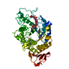
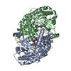
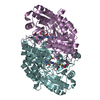
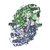
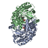
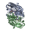
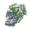
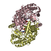
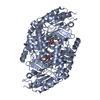
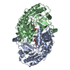
Mass: 39.098 Da / Num. of mol.: 2 / Source method: obtained synthetically / Formula: K
#3: Chemical ChemComp-PLP / PYRIDOXAL-5'-PHOSPHATE / VITAMIN B6 Phosphate


Mass: 247.142 Da / Num. of mol.: 2 / Source method: obtained synthetically / Formula: C8H10NO6P
#4: Water ChemComp-HOH / water


Mass: 18.015 Da / Num. of mol.: 556 / Source method: isolated from a natural source / Formula: H2O
Sequence detailsTRP14 IS OBSERVED AS A LEU RESIDUE.

-
Experimental details

-
Experiment

ExperimentMethod: X-RAY DIFFRACTION / Number of used crystals: 1

-
Sample preparation

CrystalDensity Matthews: 2.2 Å3/Da / Density % sol: 30 %
Crystal growpH: 6.7 / Details: pH 6.70
Crystal grow
*PLUS
pH: 7.3 / Method: vapor diffusion
Details: drop consists of equal volume of protein and precipitant solutions
Components of the solutions
*PLUS
IDConc.Common nameCrystal-IDSol-IDDetails
110 mg/mlprotein1drop
221 %PEG40001reservoirprecipitant
312 %2-propanol1reservoirprecipitant

-
Data collection

DiffractionMean temperature: 100 K
Diffraction sourceSource: SYNCHROTRON / Site: EMBL/DESY, HAMBURG / Beamline: X11 / Wavelength: 0.8265, 0.9082, 0.9918, 1.007
DetectorType: MARRESEARCH / Detector: IMAGE PLATE / Date: Oct 15, 1996
RadiationProtocol: MAD / Monochromatic (M) / Laue (L): M / Scattering type: x-ray
Radiation wavelength
IDWavelength (Å)Relative weight
10.82651
20.90821
30.99181
41.0071
ReflectionResolution: 1.8→20 Å / Num. obs: 149599 / % possible obs: 94.4 % / Redundancy: 2.2 % / Biso Wilson estimate: 17.7 Å2 / Rsym value: 0.068 / Net I/σ(I): 14
Reflection shellResolution: 1.8→1.86 Å / Mean I/σ(I) obs: 2.8 / Rsym value: 0.305 / % possible all: 81.3
Reflection
*PLUS
Num. obs: 67496 / Num. measured all: 149599 / Rmerge(I) obs: 0.068
Reflection shell
*PLUS
% possible obs: 81.3 % / Rmerge(I) obs: 0.305

-
Processing

Software
NameClassification
REFMACrefinement
DENZOdata reduction
SCALEPACKdata scaling
CCP4phasing
RefinementMethod to determine structure: MAD / Resolution: 1.8→20 Å / Cross valid method: THROUGHOUT / σ(F): 0
Details: BREAK IN CHAIN A, RESIDUE ASP 183 IS MISSING DUE TO DISORDER. ONLY MAIN-CHAIN ATOMS AND THE CB ATOM IS INCLUDED FOR GLU185 CHAIN B, DUE TO DISORDER. ONLY BACKBONE OF C-TERMINAL RESIDUE WAS ...Details: BREAK IN CHAIN A, RESIDUE ASP 183 IS MISSING DUE TO DISORDER. ONLY MAIN-CHAIN ATOMS AND THE CB ATOM IS INCLUDED FOR GLU185 CHAIN B, DUE TO DISORDER. ONLY BACKBONE OF C-TERMINAL RESIDUE WAS SEEN IN ELECTRON DENSITY
RfactorNum. reflection% reflectionSelection details
Rfree0.226 -5 %RANDOM
Rwork0.175 ---
obs-67496 94.4 %-
Displacement parametersBiso mean: 21.2 Å2
Refinement stepCycle: LAST / Resolution: 1.8→20 Å
ProteinNucleic acidLigandSolventTotal
Num. atoms6608 0 32 556 7196
Refine LS restraints
Refine-IDTypeDev idealDev ideal target
X-RAY DIFFRACTIONp_bond_d0.010.02
X-RAY DIFFRACTIONp_angle_d0.0270.04
X-RAY DIFFRACTIONp_angle_deg
X-RAY DIFFRACTIONp_planar_d0.0310.05
X-RAY DIFFRACTIONp_hb_or_metal_coord
X-RAY DIFFRACTIONp_mcbond_it1.8383
X-RAY DIFFRACTIONp_mcangle_it2.4075
X-RAY DIFFRACTIONp_scbond_it4.1646
X-RAY DIFFRACTIONp_scangle_it5.2948
X-RAY DIFFRACTIONp_plane_restr0.0203
X-RAY DIFFRACTIONp_chiral_restr
X-RAY DIFFRACTIONp_singtor_nbd0.1750.3
X-RAY DIFFRACTIONp_multtor_nbd0.260.3
X-RAY DIFFRACTIONp_xhyhbond_nbd
X-RAY DIFFRACTIONp_xyhbond_nbd
X-RAY DIFFRACTIONp_planar_tor3.57
X-RAY DIFFRACTIONp_staggered_tor16.415
X-RAY DIFFRACTIONp_orthonormal_tor
X-RAY DIFFRACTIONp_transverse_tor
X-RAY DIFFRACTIONp_special_tor15

+
About Yorodumi

-
News

-
Feb 9, 2022. New format data for meta-information of EMDB entries

New format data for meta-information of EMDB entries

  • Version 3 of the EMDB header file is now the official format.
  • The previous official version 1.9 will be removed from the archive.

Related info.:EMDB header

External links:wwPDB to switch to version 3 of the EMDB data model

-
Aug 12, 2020. Covid-19 info

Covid-19 info

URL: https://pdbjlvh1.pdbj.org/emnavi/covid19.php

New page: Covid-19 featured information page in EM Navigator.

Related info.:Covid-19 info / Mar 5, 2020. Novel coronavirus structure data

+
Mar 5, 2020. Novel coronavirus structure data

Novel coronavirus structure data

Related info.:Yorodumi Speices / Aug 12, 2020. Covid-19 info

External links:COVID-19 featured content - PDBj / Molecule of the Month (242):Coronavirus Proteases

+
Jan 31, 2019. EMDB accession codes are about to change! (news from PDBe EMDB page)

EMDB accession codes are about to change! (news from PDBe EMDB page)

  • The allocation of 4 digits for EMDB accession codes will soon come to an end. Whilst these codes will remain in use, new EMDB accession codes will include an additional digit and will expand incrementally as the available range of codes is exhausted. The current 4-digit format prefixed with “EMD-” (i.e. EMD-XXXX) will advance to a 5-digit format (i.e. EMD-XXXXX), and so on. It is currently estimated that the 4-digit codes will be depleted around Spring 2019, at which point the 5-digit format will come into force.
  • The EM Navigator/Yorodumi systems omit the EMD- prefix.

Related info.:Q: What is EMD? / ID/Accession-code notation in Yorodumi/EM Navigator

External links:EMDB Accession Codes are Changing Soon! / Contact to PDBj

+
Jul 12, 2017. Major update of PDB

Major update of PDB

  • wwPDB released updated PDB data conforming to the new PDBx/mmCIF dictionary.
  • This is a major update changing the version number from 4 to 5, and with Remediation, in which all the entries are updated.
  • In this update, many items about electron microscopy experimental information are reorganized (e.g. em_software).
  • Now, EM Navigator and Yorodumi are based on the updated data.

External links:wwPDB Remediation / Enriched Model Files Conforming to OneDep Data Standards Now Available in the PDB FTP Archive

-
Yorodumi

Thousand views of thousand structures

  • Yorodumi is a browser for structure data from EMDB, PDB, SASBDB, etc.
  • This page is also the successor to EM Navigator detail page, and also detail information page/front-end page for Omokage search.
  • The word "yorodu" (or yorozu) is an old Japanese word meaning "ten thousand". "mi" (miru) is to see.

Related info.:EMDB / PDB / SASBDB / Comparison of 3 databanks / Yorodumi Search / Aug 31, 2016. New EM Navigator & Yorodumi / Yorodumi Papers / Jmol/JSmol / Function and homology information / Changes in new EM Navigator and Yorodumi

Read more