 Movie
Movie Controller


+ Open data

- Basic information
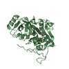
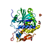
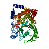
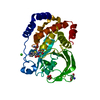
| Entry | Database: PDB / ID: 1mir | ||||||
|---|---|---|---|---|---|---|---|
| Title | RAT PROCATHEPSIN B | ||||||
 Components Components | PROCATHEPSIN B | ||||||
 Keywords Keywords |  HYDROLASE / THIOL PROTEASE / CYSTEINE PROTEASE HYDROLASE / THIOL PROTEASE / CYSTEINE PROTEASE | ||||||
| Function / homology |  Function and homology information Function and homology informationTrafficking and processing of endosomal TLR / Assembly of collagen fibrils and other multimeric structures / kininogen binding / cathepsin B / Collagen degradation / response to interleukin-4 / peptidase inhibitor complex / MHC class II antigen presentation / thyroid hormone generation / cellular response to thyroid hormone stimulus ...Trafficking and processing of endosomal TLR / Assembly of collagen fibrils and other multimeric structures / kininogen binding / cathepsin B / Collagen degradation / response to interleukin-4 / peptidase inhibitor complex / MHC class II antigen presentation / thyroid hormone generation / cellular response to thyroid hormone stimulus / proteoglycan binding / Neutrophil degranulation / response to dexamethasone / response to amine / decidualization / collagen catabolic process / response to glucose / response to mechanical stimulus / cysteine-type peptidase activity / skeletal muscle tissue development / epithelial cell differentiation / collagen binding / proteolysis involved in protein catabolic process / response to cytokine / peptide binding / protein catabolic process / sarcolemma / response to organic cyclic compound / response to peptide hormone / autophagy / cellular response to mechanical stimulus / : / melanosome / peptidase activity / spermatogenesis / neuron apoptotic process / response to ethanol / endopeptidase activity / lysosome / symbiont entry into host cell / apical plasma membrane / external side of plasma membrane / cysteine-type endopeptidase activity / protein-containing complex binding / perinuclear region of cytoplasm / cell surface / proteolysis / extracellular space / extracellular regionSimilarity search - Function | ||||||
| Biological species |  Rattus norvegicus (Norway rat) Rattus norvegicus (Norway rat) | ||||||
| Method | X-RAY DIFFRACTION / Resolution: 2.8 Å | ||||||
 Authors Authors | Cygler, M. / Sivaraman, J. / Grochulski, P. / Coulombe, R. / Storer, A.C. / Mort, J.S. | ||||||
 Citation Citation | Journal: Structure / Year: 1996 Title: Structure of rat procathepsin B: model for inhibition of cysteine protease activity by the proregion. Authors: Cygler, M. / Sivaraman, J. / Grochulski, P. / Coulombe, R. / Storer, A.C. / Mort, J.S. #1: Journal: To be PublishedTitle: Crystallization of Rat Procathepsin B Authors: Sivaraman, J. / Coulombe, R. / Magny, M.-C. / Mason, P. / Mort, J.S. / Cygler, M. | ||||||
| History |
|
- Structure visualization
| Structure viewer | Molecule: MolmilJmol/JSmol |
|---|
- Downloads & links
-Download
| PDBx/mmCIF format | 1mir.cif.gz | 149.8 KB | Display | PDBx/mmCIF format |
|---|---|---|---|---|
| PDB format | pdb1mir.ent.gz | 124.4 KB | Display | PDB format |
| PDBx/mmJSON format | 1mir.json.gz | Tree view | PDBx/mmJSON format | |
| Others |  Other downloads Other downloads |
-Validation report
| Arichive directory | https://data.pdbj.org/pub/pdb/validation_reports/mi/1mirftp://data.pdbj.org/pub/pdb/validation_reports/mi/1mir | HTTPS FTP |
|---|
-Related structure data
| Similar structure data |
|---|


-Links
 PDBj
PDBj


- Assembly
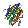
| Deposited unit | 
| ||||||||
|---|---|---|---|---|---|---|---|---|---|
| 1 | 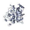
| ||||||||
| 2 | 
| ||||||||
| Unit cell |
| ||||||||
| Noncrystallographic symmetry (NCS) | NCS oper: (Code: given Matrix: (0.2123, 0.6981, -0.6839), Vector : |
-Components
| #1: Protein | Mass: 35633.625 Da / Num. of mol.: 2 / Mutation: C29S, S115A Source method: isolated from a genetically manipulated source Source: (gene. exp.) Rattus norvegicus (Norway rat) / Variant: NATURALLY OCCURRING VARIANT V223A / Production host:  Pichia pastoris (fungus) / References: UniProt: P00787, cathepsin B Pichia pastoris (fungus) / References: UniProt: P00787, cathepsin B#2: Water | ChemComp-HOH / | Water Mass: 18.015 Da / Num. of mol.: 33 / Source method: isolated from a natural source / Formula: H2O Mass: 18.015 Da / Num. of mol.: 33 / Source method: isolated from a natural source / Formula: H2O |
|---|
-Experimental details
-Experiment
| Experiment | Method: X-RAY DIFFRACTION |
|---|
- Sample preparation
| Crystal | Density Matthews: 2.84 Å3/Da / Density % sol: 56 % |
|---|---|
| Crystal | *PLUS |
| Crystal grow | *PLUS Method: unknown |
-Data collection
| Diffraction source | Wavelength: 1.5418 |
|---|---|
| Detector | Type: RIGAKU / Detector: IMAGE PLATE / Date: Jul 7, 1995 |
| Radiation | Monochromatic (M) / Laue (L): M / Scattering type: x-ray |
| Radiation wavelength | Wavelength: 1.5418 Å / Relative weight: 1 |
| Reflection | Resolution: 2.8→74 Å / Num. obs: 19015 / % possible obs: 91 % / Observed criterion σ(I): 1 / Redundancy: 4.4 % / Rmerge(I) obs: 0.097 |
- Processing
| Software |
| ||||||||||||||||||||||||||||||||||||||||||||||||||||||||||||
|---|---|---|---|---|---|---|---|---|---|---|---|---|---|---|---|---|---|---|---|---|---|---|---|---|---|---|---|---|---|---|---|---|---|---|---|---|---|---|---|---|---|---|---|---|---|---|---|---|---|---|---|---|---|---|---|---|---|---|---|---|---|
| Refinement | Resolution: 2.8→8 Å / σ(F): 2
| ||||||||||||||||||||||||||||||||||||||||||||||||||||||||||||
| Displacement parameters | Biso mean: 32.4 Å2 | ||||||||||||||||||||||||||||||||||||||||||||||||||||||||||||
| Refinement step | Cycle: LAST / Resolution: 2.8→8 Å
| ||||||||||||||||||||||||||||||||||||||||||||||||||||||||||||
| Refine LS restraints |
| ||||||||||||||||||||||||||||||||||||||||||||||||||||||||||||
| Software | *PLUS Name: X-PLOR / Version: 3.1 / Classification: refinement | ||||||||||||||||||||||||||||||||||||||||||||||||||||||||||||
| Refinement | *PLUS Rfactor all: 0.22 / Rfactor obs: 0.19 | ||||||||||||||||||||||||||||||||||||||||||||||||||||||||||||
| Solvent computation | *PLUS | ||||||||||||||||||||||||||||||||||||||||||||||||||||||||||||
| Displacement parameters | *PLUS | ||||||||||||||||||||||||||||||||||||||||||||||||||||||||||||
| Refine LS restraints | *PLUS
|
