 Movie
Movie Controller


+ Open data

- Basic information
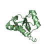
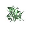
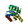
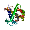
| Entry | Database: PDB / ID: 1mil | ||||||
|---|---|---|---|---|---|---|---|
| Title | TRANSFORMING PROTEIN | ||||||
 Components Components | SHC ADAPTOR PROTEIN | ||||||
 Keywords Keywords | TRANSFORMING PROTEIN / SH2 DOMAIN / PHOSPHORYLATION / COLLAGEN / GROWTH REGULATION / ALTERNATIVE INITIATION | ||||||
| Function / homology |  Function and homology information Function and homology informationregulation of superoxide metabolic process / positive regulation of cell proliferation in bone marrow / XBP1(S) activates chaperone genes / neurotrophin TRKA receptor binding / transmembrane receptor protein tyrosine kinase adaptor activity / Interleukin-15 signaling / Interleukin-2 signaling / Signaling by LTK / epidermal growth factor receptor binding / Shc-EGFR complex ...regulation of superoxide metabolic process / positive regulation of cell proliferation in bone marrow / XBP1(S) activates chaperone genes / neurotrophin TRKA receptor binding / transmembrane receptor protein tyrosine kinase adaptor activity / Interleukin-15 signaling / Interleukin-2 signaling / Signaling by LTK / epidermal growth factor receptor binding / Shc-EGFR complex / epidermal growth factor binding / Signaling by ALK / RET signaling / Interleukin-3, Interleukin-5 and GM-CSF signaling / Activated NTRK3 signals through RAS / Role of LAT2/NTAL/LAB on calcium mobilization / Activated NTRK2 signals through RAS / Interleukin receptor SHC signaling / SHC1 events in ERBB4 signaling / Signal attenuation / Signalling to RAS / SHC-related events triggered by IGF1R / SHC-mediated cascade:FGFR3 / MET activates RAS signaling / SHC-mediated cascade:FGFR2 / SHC-mediated cascade:FGFR4 / Erythropoietin activates RAS / SHC-mediated cascade:FGFR1 / ephrin receptor binding / Signaling by CSF3 (G-CSF) / insulin-like growth factor receptor binding / Tie2 Signaling / phosphotyrosine residue binding / Integrin signaling / SHC1 events in EGFR signaling / FCERI mediated Ca+2 mobilization / insulin-like growth factor receptor signaling pathway / Insulin receptor signalling cascade / SHC1 events in ERBB2 signaling / Constitutive Signaling by Overexpressed ERBB2 / negative regulation of angiogenesis / insulin receptor binding / FCERI mediated MAPK activation / Signaling by ERBB2 TMD/JMD mutants / cell-cell adhesion / Constitutive Signaling by EGFRvIII / Signaling by ERBB2 ECD mutants / Signaling by ERBB2 KD Mutants / receptor tyrosine kinase binding / phospholipid binding / cellular response to growth factor stimulus / epidermal growth factor receptor signaling pathway / Signaling by CSF1 (M-CSF) in myeloid cells / Signaling by ALK fusions and activated point mutants / insulin receptor signaling pathway / DAP12 signaling / GPER1 signaling / Constitutive Signaling by Ligand-Responsive EGFR Cancer Variants / heart development / RAF/MAP kinase cascade / actin cytoskeleton organization / angiogenesis / Extra-nuclear estrogen signaling / positive regulation of ERK1 and ERK2 cascade / positive regulation of MAPK cascade / defense response to bacterium / intracellular signal transduction / mitochondrial matrix / focal adhesion / negative regulation of DNA-templated transcription / positive regulation of cell population proliferation / negative regulation of apoptotic process / positive regulation of DNA-templated transcription / plasma membrane / cytosol Similarity search - Function | ||||||
| Biological species |  Homo sapiens (human) Homo sapiens (human) | ||||||
| Method |  X-RAY DIFFRACTION / Resolution: 2.7 Å X-RAY DIFFRACTION / Resolution: 2.7 Å | ||||||
 Authors Authors | Mikol, V. | ||||||
 Citation Citation | Journal: J.Mol.Biol. / Year: 1995 Title: Crystal structure of the SH2 domain from the adaptor protein SHC: a model for peptide binding based on X-ray and NMR data. Authors: Mikol, V. / Baumann, G. / Zurini, M.G. / Hommel, U. #1: Journal: Acta Crystallogr.,Sect.D / Year: 1994Title: Crystallization of the Complex between Cyclophilin a and Cyclosporin Derivatives: The Use of Cross-Seeding Authors: Mikol, V. / Duc, D. #2: Journal: J.Mol.Biol. / Year: 1993Title: X-Ray Structure of a Monomeric Cyclophilin A-Cyclosporin a Crystal Complex at 2.1 A Resolution Authors: Mikol, V. / Kallen, J. / Pflugl, G. / Walkinshaw, M.D. #3: Journal: Cell(Cambridge,Mass.) / Year: 1992Title: A Novel Transforming Protein (Shc) with an Sh2 Domain is Implicated in Mitogenic Signal Transduction Authors: Pelicci, G. / Lanfrancone, L. / Grignani, F. / Mcglade, J. / Cavallo, F. / Forni, G. / Nicoletti, I. / Grignani, F. / Pawson, T. / Pelicci, P.G. | ||||||
| History |
|
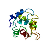
- Structure visualization
| Structure viewer | Molecule: MolmilJmol/JSmol |
|---|
- Downloads & links
-Download
| PDBx/mmCIF format | 1mil.cif.gz | 34 KB | Display | PDBx/mmCIF format |
|---|---|---|---|---|
| PDB format | pdb1mil.ent.gz | 22.6 KB | Display | PDB format |
| PDBx/mmJSON format | 1mil.json.gz | Tree view | PDBx/mmJSON format | |
| Others |  Other downloads Other downloads |
-Validation report
| Arichive directory | https://data.pdbj.org/pub/pdb/validation_reports/mi/1milftp://data.pdbj.org/pub/pdb/validation_reports/mi/1mil | HTTPS FTP |
|---|
-Related structure data
| Similar structure data |
|---|


-Links
 PDBj
PDBj


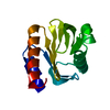
- Assembly
| Deposited unit | 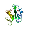
| ||||||||
|---|---|---|---|---|---|---|---|---|---|
| 1 |
| ||||||||
| Unit cell |
|
-Components
| #1: Protein | Mass: 11713.277 Da / Num. of mol.: 1 / Fragment: PHOSPHOTYROSINE RECOGNITION DOMAIN SH2 Source method: isolated from a genetically manipulated source Source: (gene. exp.) Homo sapiens (human) / Gene: T7 / Plasmid: T7 / Gene (production host): T7 / Production host:  | ||
|---|---|---|---|
| #2: Water | ChemComp-HOH /  Mass: 18.015 Da / Num. of mol.: 85 / Source method: isolated from a natural source / Formula: H2O Mass: 18.015 Da / Num. of mol.: 85 / Source method: isolated from a natural source / Formula: H2O | ||
| Compound details | RESIDUE ARG 16 OF SHC ADOPTS ALTERNATE CONFORMATI| Sequence details | RESIDUES 1 AND 2 CORRESPOND TO RESIDUES OF THE T7 GENE LEADER SEQUENCE REQUIRED FOR EXPRESSION AND ...RESIDUES 1 AND 2 CORRESPOND | |
-Experimental details
-Experiment
| Experiment | Method: X-RAY DIFFRACTION |
|---|
- Sample preparation
| Crystal | Density Matthews: 2.28 Å3/Da / Density % sol: 46.05 % | ||||||||||||||||||||||||||||||||||||||||||||||||||||||
|---|---|---|---|---|---|---|---|---|---|---|---|---|---|---|---|---|---|---|---|---|---|---|---|---|---|---|---|---|---|---|---|---|---|---|---|---|---|---|---|---|---|---|---|---|---|---|---|---|---|---|---|---|---|---|---|
| Crystal | *PLUS Density % sol: 46.2 % | ||||||||||||||||||||||||||||||||||||||||||||||||||||||
| Crystal grow | *PLUS Temperature: 22 ℃ / pH: 5 / Method: vapor diffusion, hanging drop | ||||||||||||||||||||||||||||||||||||||||||||||||||||||
| Components of the solutions | *PLUS
|
-Data collection
| Radiation | Scattering type: x-ray |
|---|---|
| Radiation wavelength | Relative weight: 1 |
| Reflection | Resolution: 2.7→10 Å / Num. obs: 3028 / % possible obs: 94 % / Observed criterion σ(I): 2 |
| Reflection | *PLUS Num. measured all: 7344 / Rmerge(I) obs: 0.051 |
- Processing
| Software |
| ||||||||||||||||||||||||||||||||||||||||||||||||||||||||||||
|---|---|---|---|---|---|---|---|---|---|---|---|---|---|---|---|---|---|---|---|---|---|---|---|---|---|---|---|---|---|---|---|---|---|---|---|---|---|---|---|---|---|---|---|---|---|---|---|---|---|---|---|---|---|---|---|---|---|---|---|---|---|
| Refinement | Resolution: 2.7→8 Å / σ(F): 2 /
| ||||||||||||||||||||||||||||||||||||||||||||||||||||||||||||
| Refinement step | Cycle: LAST / Resolution: 2.7→8 Å
| ||||||||||||||||||||||||||||||||||||||||||||||||||||||||||||
| Refine LS restraints |
| ||||||||||||||||||||||||||||||||||||||||||||||||||||||||||||
| Refinement | *PLUS | ||||||||||||||||||||||||||||||||||||||||||||||||||||||||||||
| Solvent computation | *PLUS | ||||||||||||||||||||||||||||||||||||||||||||||||||||||||||||
| Displacement parameters | *PLUS |
