 Movie
Movie Controller


+ Open data

- Basic information
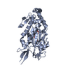
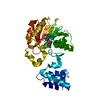
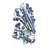
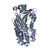
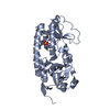
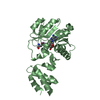
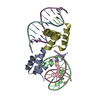
| Entry | Database: PDB / ID: 1m93 | ||||||
|---|---|---|---|---|---|---|---|
| Title | 1.65 A Structure of Cleaved Viral Serpin CRMA | ||||||
 Components Components | (Serine proteinase inhibitor 2 ) x 3 ) x 3 | ||||||
 Keywords Keywords | VIRAL PROTEIN / SERPIN / CRMA / APOPTOSIS / ICE INHIBITOR | ||||||
| Function / homology |  Function and homology information Function and homology information: / Microbial modulation of RIPK1-mediated regulated necrosis / cysteine-type endopeptidase inhibitor activity / protein sequestering activity / Regulation of TNFR1 signaling / serine-type endopeptidase inhibitor activity / : / symbiont-mediated suppression of host cytoplasmic pattern recognition receptor signaling pathway via inhibition of TBK1 activity / host cell cytoplasm / extracellular space / cytoplasmSimilarity search - Function | ||||||
| Biological species |  Cowpox virus Cowpox virus | ||||||
| Method | X-RAY DIFFRACTION / SYNCHROTRON / MOLECULAR REPLACEMENT / Resolution: 1.65 Å | ||||||
 Authors Authors | Simonovic, M. / Gettins, P.G.W. / Volz, K. | ||||||
 Citation Citation | Journal: Protein Sci. / Year: 2000 Title: Crystal structure of viral serpin crmA provides insights into its mechanism of cysteine proteinase inhibition Authors: Simonovic, M. / Gettins, P.G.W. / Volz, K. #1: Journal: Acta Crystallogr.,Sect.D / Year: 2000Title: Crystallization and preliminary X-ray diffraction analysis of a recombinant cysteine-free mutant of crmA Authors: Simonovic, M. / Gettins, P.G.W. / Volz, K. | ||||||
| History |
|
- Structure visualization
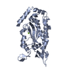
| Structure viewer | Molecule: MolmilJmol/JSmol |
|---|
- Downloads & links
-Download
| PDBx/mmCIF format | 1m93.cif.gz | 80.6 KB | Display | PDBx/mmCIF format |
|---|---|---|---|---|
| PDB format | pdb1m93.ent.gz | 60.1 KB | Display | PDB format |
| PDBx/mmJSON format | 1m93.json.gz | Tree view | PDBx/mmJSON format | |
| Others |  Other downloads Other downloads |
-Validation report
| Arichive directory | https://data.pdbj.org/pub/pdb/validation_reports/m9/1m93ftp://data.pdbj.org/pub/pdb/validation_reports/m9/1m93 | HTTPS FTP |
|---|
-Related structure data
| Related structure data |  1c8oSC S: Starting model for refinement C: citing same article ( |
|---|---|
| Similar structure data |


-Links
 PDBj
PDBj


- Assembly
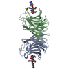
| Deposited unit | 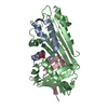
| ||||||||
|---|---|---|---|---|---|---|---|---|---|
| 1 |
| ||||||||
| Unit cell |
|
-Components
| #1: Protein | / cytokine response modifier protein A / Serpin 2 / Serp-2 / ICE inhibitor / Hemorrhage-inducing 38 kDa protein Mass: 6079.818 Da / Num. of mol.: 1 / Fragment: RESIDUES 1-55 Source method: isolated from a genetically manipulated source Source: (gene. exp.) Cowpox virus / Genus: Orthopoxvirus / Gene: crmA / Plasmid: pQE-60 / Production host:  Escherichia coli (E. coli) / Strain (production host): SG13009 / References: UniProt: P07385 Escherichia coli (E. coli) / Strain (production host): SG13009 / References: UniProt: P07385 |
|---|---|
| #2: Protein | / cytokine response modifier protein A / Serpin 2 / Serp-2 / ICE inhibitor / Hemorrhage-inducing 38 kDa protein Mass: 27403.771 Da / Num. of mol.: 1 / Fragment: RESIDUES 56-300 / Mutation: C93S, C102S, C124S, C223S, C269S, C298S Source method: isolated from a genetically manipulated source Source: (gene. exp.) Cowpox virus / Genus: Orthopoxvirus / Gene: crmA / Plasmid: pQE-60 / Production host: Escherichia coli (E. coli) / Strain (production host): SG13009 / References: UniProt: P07385 |
| #3: Protein/peptide | / cytokine response modifier protein A / Serpin 2 / Serp-2 / ICE inhibitor / Hemorrhage-inducing 38 kDa protein Mass: 4517.984 Da / Num. of mol.: 1 / Fragment: RESIDUES 301-341 / Mutation: C304S, C313S, C336S Source method: isolated from a genetically manipulated source Source: (gene. exp.) Cowpox virus / Genus: Orthopoxvirus / Gene: crmA / Plasmid: pQE-60 / Production host: Escherichia coli (E. coli) / Strain (production host): SG13009 / References: UniProt: P07385 |
| #4: Chemical | ChemComp-PO4 / Phosphate  Mass: 94.971 Da / Num. of mol.: 1 / Source method: obtained synthetically / Formula: PO4 Mass: 94.971 Da / Num. of mol.: 1 / Source method: obtained synthetically / Formula: PO4 |
| #5: Water | ChemComp-HOH / Water Mass: 18.015 Da / Num. of mol.: 146 / Source method: isolated from a natural source / Formula: H2O Mass: 18.015 Da / Num. of mol.: 146 / Source method: isolated from a natural source / Formula: H2O |
-Experimental details
-Experiment
| Experiment | Method: X-RAY DIFFRACTION / Number of used crystals: 1 |
|---|
- Sample preparation
| Crystal | Density Matthews: 2.76 Å3/Da / Density % sol: 59.46 % |
|---|---|
| Crystal grow | Temperature: 293 K / Method: vapor diffusion, hanging drop / pH: 6.5 Details: sodium/potassium phosphate 1.6M, pH 6.5, VAPOR DIFFUSION, HANGING DROP, temperature 293K |
-Data collection
| Diffraction | Mean temperature: 100 K |
|---|---|
| Diffraction source | Source: SYNCHROTRON / Site: APS  / Beamline: 14-BM-C / Wavelength: 1 / Beamline: 14-BM-C / Wavelength: 1 |
| Detector | Type: ADSC QUANTUM 4 / Detector: CCD / Date: Feb 12, 2001 |
| Radiation | Protocol: SINGLE WAVELENGTH / Monochromatic (M) / Laue (L): M / Scattering type: x-ray |
| Radiation wavelength | Wavelength: 1 Å / Relative weight: 1 |
| Reflection | Resolution: 1.65→31.54 Å / Num. all: 54375 / Num. obs: 53225 / % possible obs: 94.2 % / Observed criterion σ(F): 0 / Observed criterion σ(I): 0 / Redundancy: 5.6 % / Rmerge(I) obs: 0.039 / Net I/σ(I): 32.6 |
| Reflection shell | Resolution: 1.65→1.71 Å / Redundancy: 2.8 % / Rmerge(I) obs: 0.47 / Mean I/σ(I) obs: 2.1 / % possible all: 83.5 |
- Processing
| Software |
| |||||||||||||||||||||||||||||||||
|---|---|---|---|---|---|---|---|---|---|---|---|---|---|---|---|---|---|---|---|---|---|---|---|---|---|---|---|---|---|---|---|---|---|---|
| Refinement | Method to determine structure: MOLECULAR REPLACEMENT Starting model: PDB ID 1C8O Resolution: 1.65→31.54 Å / Num. parameters: 25438 / Num. restraintsaints: 31394 / Cross valid method: FREE R / σ(F): 0 / Stereochemistry target values: ENGH AND HUBER Details: ANISOTROPIC SCALING APPLIED BY THE METHOD OF PARKIN, MOEZZI & HOPE, J.APPL.CRYST.28(1995)53-56 ANISOTROPIC REFINEMENT REDUCED FREE R (NO CUTOFF) BY ?. Following side-chains are disordered: ...Details: ANISOTROPIC SCALING APPLIED BY THE METHOD OF PARKIN, MOEZZI & HOPE, J.APPL.CRYST.28(1995)53-56 ANISOTROPIC REFINEMENT REDUCED FREE R (NO CUTOFF) BY ?. Following side-chains are disordered: ILE57, and ASP122 Following amino-acids are missing: 47 GLU LYS GLU ALA ASP LYS ASN LYS ASP 55; 301 VAL ALA ASP SER ALA SER THR VAL 308
| |||||||||||||||||||||||||||||||||
| Solvent computation | Solvent model: MOEWS & KRETSINGER, J.MOL.BIOL.91(1973)201-228 | |||||||||||||||||||||||||||||||||
| Refine analyze | Num. disordered residues: 18 / Occupancy sum hydrogen: 0 / Occupancy sum non hydrogen: 2777 | |||||||||||||||||||||||||||||||||
| Refinement step | Cycle: LAST / Resolution: 1.65→31.54 Å
| |||||||||||||||||||||||||||||||||
| Refine LS restraints |
|
