 Movie
Movie Controller

[English] 日本語
 Yorodumi
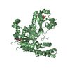
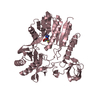
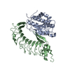
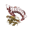
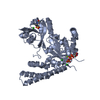
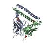
- PDB-1m10: Crystal structure of the complex of Glycoprotein Ib alpha and the... -
+ Open data

- Basic information
| Entry | Database: PDB / ID: 1m10 | ||||||
|---|---|---|---|---|---|---|---|
| Title | Crystal structure of the complex of Glycoprotein Ib alpha and the von Willebrand Factor A1 Domain | ||||||
 Components Components |
| ||||||
 Keywords Keywords | BLOOD CLOTTING / leucine-rich repeat / HEMOSTASIS / DINUCLEOTIDE BINDING FOLD | ||||||
| Function / homology |  Function and homology information Function and homology informationthrombin-activated receptor activity / glycoprotein Ib-IX-V complex / Defective VWF binding to collagen type I / Enhanced cleavage of VWF variant by ADAMTS13 / Defective VWF cleavage by ADAMTS13 variant / Defective F8 binding to von Willebrand factor / Enhanced binding of GP1BA variant to VWF multimer:collagen / Defective binding of VWF variant to GPIb:IX:V / Weibel-Palade body / blood coagulation, intrinsic pathway ...thrombin-activated receptor activity / glycoprotein Ib-IX-V complex / Defective VWF binding to collagen type I / Enhanced cleavage of VWF variant by ADAMTS13 / Defective VWF cleavage by ADAMTS13 variant / Defective F8 binding to von Willebrand factor / Enhanced binding of GP1BA variant to VWF multimer:collagen / Defective binding of VWF variant to GPIb:IX:V / Weibel-Palade body / blood coagulation, intrinsic pathway / hemostasis / Defective F9 activation / platelet alpha granule / Platelet Adhesion to exposed collagen / positive regulation of platelet activation / megakaryocyte development / GP1b-IX-V activation signalling / p130Cas linkage to MAPK signaling for integrins / regulation of blood coagulation / Defective F8 cleavage by thrombin / Platelet Aggregation (Plug Formation) / cell-substrate adhesion / GRB2:SOS provides linkage to MAPK signaling for Integrins / positive regulation of intracellular signal transduction / immunoglobulin binding / Integrin cell surface interactions / fibrinolysis / collagen binding / Intrinsic Pathway of Fibrin Clot Formation / Integrin signaling / release of sequestered calcium ion into cytosol / platelet alpha granule lumen / Signaling by high-kinase activity BRAF mutants / RUNX1 regulates genes involved in megakaryocyte differentiation and platelet function / MAP2K and MAPK activation / platelet activation / response to wounding / extracellular matrix / integrin binding / : / cell morphogenesis / blood coagulation / Signaling by RAF1 mutants / Signaling by moderate kinase activity BRAF mutants / Paradoxical activation of RAF signaling by kinase inactive BRAF / Signaling downstream of RAS mutants / Signaling by BRAF and RAF1 fusions / Platelet degranulation / protein-folding chaperone binding / protease binding / cell surface receptor signaling pathway / cell adhesion / external side of plasma membrane / cell surface / endoplasmic reticulum / extracellular space / extracellular exosome / extracellular region / identical protein binding / membrane / plasma membrane Similarity search - Function | ||||||
| Biological species |  Homo sapiens (human) Homo sapiens (human) | ||||||
| Method |  X-RAY DIFFRACTION / SYNCHROTRON / MOLECULAR REPLACEMENT / Resolution: 3.1 Å X-RAY DIFFRACTION / SYNCHROTRON / MOLECULAR REPLACEMENT / Resolution: 3.1 Å | ||||||
 Authors Authors | Huizinga, E.G. / Tsuji, S. / Romijn, R.A.P. / Schiphorst, M.E. / de Groot, P.G. / Sixma, J.J. / Gros, P. | ||||||
 Citation Citation | Journal: Science / Year: 2002 Title: Structures of glycoprotein Ibalpha and its complex with von Willebrand factor A1 domain. Authors: Huizinga, E.G. / Tsuji, S. / Romijn, R.A. / Schiphorst, M.E. / de Groot, P.G. / Sixma, J.J. / Gros, P. | ||||||
| History |
|
- Structure visualization
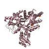
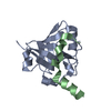
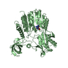
| Structure viewer | Molecule: MolmilJmol/JSmol |
|---|
- Downloads & links
-Download
| PDBx/mmCIF format | 1m10.cif.gz | 102.9 KB | Display | PDBx/mmCIF format |
|---|---|---|---|---|
| PDB format | pdb1m10.ent.gz | 79.4 KB | Display | PDB format |
| PDBx/mmJSON format | 1m10.json.gz | Tree view | PDBx/mmJSON format | |
| Others |  Other downloads Other downloads |
-Validation report
| Arichive directory | https://data.pdbj.org/pub/pdb/validation_reports/m1/1m10ftp://data.pdbj.org/pub/pdb/validation_reports/m1/1m10 | HTTPS FTP |
|---|
-Related structure data
| Related structure data | 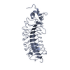 1m0zSC  1auqS S: Starting model for refinement C: citing same article ( |
|---|---|
| Similar structure data |

-Links
 PDBj
PDBj


- Assembly
| Deposited unit | 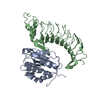
| ||||||||
|---|---|---|---|---|---|---|---|---|---|
| 1 |
| ||||||||
| Unit cell |
|
-Components
| #1: Protein | Mass: 23731.447 Da / Num. of mol.: 1 / Fragment: A1 domain / Mutation: R543Q Source method: isolated from a genetically manipulated source Source: (gene. exp.) Homo sapiens (human) / Gene: VWF / Plasmid: pPIC9 / Production host:  Pichia pastoris (fungus) / Strain (production host): GS115 / References: UniProt: P04275 Pichia pastoris (fungus) / Strain (production host): GS115 / References: UniProt: P04275 |
|---|---|
| #2: Protein | Mass: 32334.842 Da / Num. of mol.: 1 / Fragment: von Willebrand Factor binding domain / Mutation: N21Q N159Q M239V Source method: isolated from a genetically manipulated source Source: (gene. exp.) Homo sapiens (human) / Gene: GP1BA / Plasmid: pCDNA3.1 / Production host:  Mesocricetus auratus (golden hamster) / Tissue (production host): kidney / References: UniProt: P07359 Mesocricetus auratus (golden hamster) / Tissue (production host): kidney / References: UniProt: P07359 |
| Has protein modification | Y |
-Experimental details
-Experiment
| Experiment | Method: X-RAY DIFFRACTION / Number of used crystals: 1 |
|---|
- Sample preparation
| Crystal | Density Matthews: 2.59 Å3/Da / Density % sol: 52.47 % | |||||||||||||||||||||||||||||||||||||||||||||||||
|---|---|---|---|---|---|---|---|---|---|---|---|---|---|---|---|---|---|---|---|---|---|---|---|---|---|---|---|---|---|---|---|---|---|---|---|---|---|---|---|---|---|---|---|---|---|---|---|---|---|---|
| Crystal grow | Temperature: 277 K / Method: vapor diffusion, hanging drop / pH: 5.5 Details: PEG 3000, sodium chloride, MES, pH 5.5, VAPOR DIFFUSION, HANGING DROP, temperature 277K | |||||||||||||||||||||||||||||||||||||||||||||||||
| Crystal grow | *PLUS Temperature: 4 ℃ / pH: 7.8 | |||||||||||||||||||||||||||||||||||||||||||||||||
| Components of the solutions | *PLUS
|
-Data collection
| Diffraction | Mean temperature: 100 K |
|---|---|
| Diffraction source | Source: SYNCHROTRON / Site: EMBL/DESY, HAMBURG  / Beamline: X11 / Wavelength: 0.8075 Å / Beamline: X11 / Wavelength: 0.8075 Å |
| Detector | Type: MARRESEARCH / Detector: CCD / Date: Dec 12, 2001 |
| Radiation | Protocol: SINGLE WAVELENGTH / Monochromatic (M) / Laue (L): M / Scattering type: x-ray |
| Radiation wavelength | Wavelength: 0.8075 Å / Relative weight: 1 |
| Reflection | Resolution: 3.09→30.5 Å / Num. all: 10454 / Num. obs: 10454 / % possible obs: 99.9 % / Observed criterion σ(I): -3.7 / Redundancy: 5.8 % / Rmerge(I) obs: 0.087 / Net I/σ(I): 19.3 |
| Reflection shell | Resolution: 3.09→3.2 Å / Redundancy: 5.4 % / Rmerge(I) obs: 0.48 / Mean I/σ(I) obs: 3.6 / Num. unique all: 1037 / % possible all: 99.9 |
| Reflection | *PLUS Highest resolution: 3.1 Å / Lowest resolution: 40 Å |
| Reflection shell | *PLUS Highest resolution: 3.1 Å / % possible obs: 99.9 % / Rmerge(I) obs: 0.48 |
- Processing
| Software |
| ||||||||||||||||||||||||||||||||||||
|---|---|---|---|---|---|---|---|---|---|---|---|---|---|---|---|---|---|---|---|---|---|---|---|---|---|---|---|---|---|---|---|---|---|---|---|---|---|
| Refinement | Method to determine structure: MOLECULAR REPLACEMENT Starting model: PDB entries 1AUQ and 1M0Z Resolution: 3.1→30.5 Å / Rfactor Rfree error: 0.013 / Data cutoff high absF: 1123736.97 / Data cutoff low absF: 0 / Isotropic thermal model: ISOTROPIC RESTRAINED / Cross valid method: THROUGHOUT / σ(F): 0 / Stereochemistry target values: Engh & Huber
| ||||||||||||||||||||||||||||||||||||
| Solvent computation | Solvent model: FLAT MODEL / Bsol: 20.2789 Å2 / ksol: 0.300589 e/Å3 | ||||||||||||||||||||||||||||||||||||
| Displacement parameters | Biso mean: 52.2 Å2
| ||||||||||||||||||||||||||||||||||||
| Refine analyze |
| ||||||||||||||||||||||||||||||||||||
| Refinement step | Cycle: LAST / Resolution: 3.1→30.5 Å
| ||||||||||||||||||||||||||||||||||||
| Refine LS restraints |
| ||||||||||||||||||||||||||||||||||||
| LS refinement shell | Resolution: 3.1→3.29 Å / Rfactor Rfree error: 0.04 / Total num. of bins used: 6
| ||||||||||||||||||||||||||||||||||||
| Xplor file |
| ||||||||||||||||||||||||||||||||||||
| Refinement | *PLUS | ||||||||||||||||||||||||||||||||||||
| Solvent computation | *PLUS | ||||||||||||||||||||||||||||||||||||
| Displacement parameters | *PLUS | ||||||||||||||||||||||||||||||||||||
| Refine LS restraints | *PLUS
|
