 Movie
Movie Controller

[English] 日本語
 Yorodumi
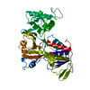
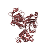
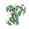
- PDB-1lv0: Crystal structure of the Rab effector guanine nucleotide dissocia... -
+ Open data

- Basic information
| Entry | Database: PDB / ID: 1lv0 | ||||||
|---|---|---|---|---|---|---|---|
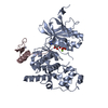
| Title | Crystal structure of the Rab effector guanine nucleotide dissociation inhibitor (GDI) in complex with a geranylgeranyl (GG) peptide | ||||||
 Components Components | RAB GDP disossociation inhibitor alpha | ||||||
 Keywords Keywords | SIGNALING PROTEIN / Protein-ligand complex | ||||||
| Function / homology |  Function and homology information Function and homology informationRAB GEFs exchange GTP for GDP on RABs / Rab GDP-dissociation inhibitor activity / negative regulation of protein targeting to membrane / Rab protein signal transduction / negative regulation of axonogenesis / vesicle-mediated transport / GTPase activator activity / protein transport / Golgi apparatus / cytosol Similarity search - Function | ||||||
| Biological species |  | ||||||
| Method |  X-RAY DIFFRACTION / SYNCHROTRON / MOLECULAR REPLACEMENT / Resolution: 2 Å X-RAY DIFFRACTION / SYNCHROTRON / MOLECULAR REPLACEMENT / Resolution: 2 Å | ||||||
 Authors Authors | An, Y. / Shao, Y. / Alory, C. / Matteson, J. / Sakisaka, T. / Chen, W. / Gibbs, R.A. / Wilson, I.A. / Balch, W.E. | ||||||
 Citation Citation | Journal: Structure / Year: 2003 Title: Geranylgeranyl switching regulates GDI-Rab GTPase recycling. Authors: An, Y. / Shao, Y. / Alory, C. / Matteson, J. / Sakisaka, T. / Chen, W. / Gibbs, R.A. / Wilson, I.A. / Balch, W.E. | ||||||
| History |
|
- Structure visualization
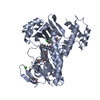
| Structure viewer | Molecule: MolmilJmol/JSmol |
|---|
- Downloads & links
-Download
| PDBx/mmCIF format | 1lv0.cif.gz | 108.7 KB | Display | PDBx/mmCIF format |
|---|---|---|---|---|
| PDB format | pdb1lv0.ent.gz | 81.3 KB | Display | PDB format |
| PDBx/mmJSON format | 1lv0.json.gz | Tree view | PDBx/mmJSON format | |
| Others |  Other downloads Other downloads |
-Validation report
| Arichive directory | https://data.pdbj.org/pub/pdb/validation_reports/lv/1lv0ftp://data.pdbj.org/pub/pdb/validation_reports/lv/1lv0 | HTTPS FTP |
|---|
-Related structure data
| Related structure data | 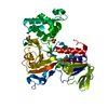 1d5tS S: Starting model for refinement |
|---|---|
| Similar structure data |


-Links
 PDBj
PDBj- Assembly
| Deposited unit | 
| ||||||||
|---|---|---|---|---|---|---|---|---|---|
| 1 |
| ||||||||
| Unit cell |
|
-Components
| #1: Protein | Mass: 50896.754 Da / Num. of mol.: 1 Source method: isolated from a genetically manipulated source Source: (gene. exp.)  | ||||
|---|---|---|---|---|---|
| #2: Chemical |   Mass: 96.063 Da / Num. of mol.: 3 / Source method: obtained synthetically / Formula: SO4 Mass: 96.063 Da / Num. of mol.: 3 / Source method: obtained synthetically / Formula: SO4#3: Chemical | ChemComp-GER / | 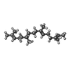  Mass: 274.484 Da / Num. of mol.: 1 / Source method: obtained synthetically / Formula: C20H34 Mass: 274.484 Da / Num. of mol.: 1 / Source method: obtained synthetically / Formula: C20H34#4: Water | ChemComp-HOH / |  Mass: 18.015 Da / Num. of mol.: 368 / Source method: isolated from a natural source / Formula: H2O Mass: 18.015 Da / Num. of mol.: 368 / Source method: isolated from a natural source / Formula: H2O |
-Experimental details
-Experiment
| Experiment | Method: X-RAY DIFFRACTION / Number of used crystals: 1 |
|---|
- Sample preparation
| Crystal | Density Matthews: 2.27 Å3/Da / Density % sol: 45.79 % |
|---|---|
| Crystal grow | Temperature: 296 K / Method: vapor diffusion, sitting drop / pH: 6.2 Details: Ammonium Sulfate, pH 6.2, VAPOR DIFFUSION, SITTING DROP, temperature 296K |
| Crystal grow | *PLUS Temperature: 22.5 ℃ / pH: 6.25 / Method: vapor diffusion, sitting dropDetails: Luan, P., (2000) Traffic, 1, 270., Schalk, I., (1996) Nature,381, 42. |
| Components of the solutions | *PLUS Conc.: 1.73 M / Common name: ammonium sulfate |
-Data collection
| Diffraction | Mean temperature: 98 K |
|---|---|
| Diffraction source | Source: SYNCHROTRON / Site: SSRL  / Beamline: BL9-2 / Wavelength: 1.033 Å / Beamline: BL9-2 / Wavelength: 1.033 Å |
| Detector | Type: ADSC QUANTUM 4 / Detector: CCD / Date: Feb 12, 2000 |
| Radiation | Monochromator: Mirror / Protocol: SINGLE WAVELENGTH / Monochromatic (M) / Laue (L): M / Scattering type: x-ray |
| Radiation wavelength | Wavelength: 1.033 Å / Relative weight: 1 |
| Reflection | Resolution: 2→20 Å / Num. all: 30213 / Num. obs: 30213 / % possible obs: 95.7 % / Observed criterion σ(F): 0 / Observed criterion σ(I): 0 / Redundancy: 2.3 % / Biso Wilson estimate: 12.7 Å2 / Rmerge(I) obs: 0.061 / Rsym value: 0.061 / Net I/σ(I): 12.7 |
| Reflection shell | Resolution: 2→2.03 Å / Rmerge(I) obs: 0.419 / Mean I/σ(I) obs: 1.4 / Num. unique all: 1325 / Rsym value: 0.419 / % possible all: 85.9 |
| Reflection | *PLUS Num. measured all: 68840 |
| Reflection shell | *PLUS % possible obs: 85.9 % |
- Processing
| Software |
| ||||||||||||||||||||||||||||||||||||
|---|---|---|---|---|---|---|---|---|---|---|---|---|---|---|---|---|---|---|---|---|---|---|---|---|---|---|---|---|---|---|---|---|---|---|---|---|---|
| Refinement | Method to determine structure: MOLECULAR REPLACEMENT Starting model: PDB ENTRY 1D5T Resolution: 2→19.96 Å / Rfactor Rfree error: 0.005 / Isotropic thermal model: RESTRAINED / Cross valid method: THROUGHOUT / σ(F): 0 / Stereochemistry target values: Engh & Huber
| ||||||||||||||||||||||||||||||||||||
| Solvent computation | Solvent model: FLAT MODEL / Bsol: 54.4971 Å2 / ksol: 0.342343 e/Å3 | ||||||||||||||||||||||||||||||||||||
| Displacement parameters | Biso mean: 29.2 Å2
| ||||||||||||||||||||||||||||||||||||
| Refine analyze | Luzzati coordinate error free: 0.3 Å / Luzzati sigma a free: 0.32 Å | ||||||||||||||||||||||||||||||||||||
| Refinement step | Cycle: LAST / Resolution: 2→19.96 Å
| ||||||||||||||||||||||||||||||||||||
| Refine LS restraints |
| ||||||||||||||||||||||||||||||||||||
| LS refinement shell | Resolution: 2→2.03 Å / Rfactor Rfree error: 0.032 / Total num. of bins used: 20
| ||||||||||||||||||||||||||||||||||||
| Xplor file |
| ||||||||||||||||||||||||||||||||||||
| Refinement | *PLUS Lowest resolution: 20 Å / % reflection Rfree: 5 % | ||||||||||||||||||||||||||||||||||||
| Solvent computation | *PLUS | ||||||||||||||||||||||||||||||||||||
| Displacement parameters | *PLUS | ||||||||||||||||||||||||||||||||||||
| Refine LS restraints | *PLUS
|
