 Movie
Movie Controller

[English] 日本語
 Yorodumi
- PDB-1luf: Crystal Structure of the MuSK Tyrosine Kinase: Insights into Rece... -
+ Open data

- Basic information
| Entry | Database: PDB / ID: 1luf | ||||||
|---|---|---|---|---|---|---|---|
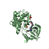
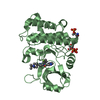
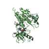
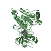
| Title | Crystal Structure of the MuSK Tyrosine Kinase: Insights into Receptor Autoregulation | ||||||
 Components Components | muscle-specific tyrosine kinase receptor musk | ||||||
 Keywords Keywords | TRANSFERASE / phosphorylation / signal transduction / mass spectrometry | ||||||
| Function / homology |  Function and homology information Function and homology informationpositive regulation of synaptic assembly at neuromuscular junction / regulation of synaptic assembly at neuromuscular junction / positive regulation of motor neuron apoptotic process / skeletal muscle acetylcholine-gated channel clustering / positive regulation of synaptic transmission, cholinergic / Wnt-protein binding / positive regulation of skeletal muscle acetylcholine-gated channel clustering / enzyme-linked receptor protein signaling pathway / neurotransmitter receptor localization to postsynaptic specialization membrane / response to muscle activity ...positive regulation of synaptic assembly at neuromuscular junction / regulation of synaptic assembly at neuromuscular junction / positive regulation of motor neuron apoptotic process / skeletal muscle acetylcholine-gated channel clustering / positive regulation of synaptic transmission, cholinergic / Wnt-protein binding / positive regulation of skeletal muscle acetylcholine-gated channel clustering / enzyme-linked receptor protein signaling pathway / neurotransmitter receptor localization to postsynaptic specialization membrane / response to muscle activity / receptor clustering / neuromuscular junction development / positive regulation of Rac protein signal transduction / cochlea development / response to electrical stimulus / response to axon injury / transmembrane receptor protein tyrosine kinase activity / cell surface receptor protein tyrosine kinase signaling pathway / cell projection / PDZ domain binding / neuromuscular junction / receptor protein-tyrosine kinase / memory / long-term synaptic potentiation / retina development in camera-type eye / protein tyrosine kinase activity / postsynaptic membrane / cell differentiation / receptor complex / negative regulation of gene expression / external side of plasma membrane / synapse / positive regulation of gene expression / regulation of DNA-templated transcription / protein kinase binding / ATP binding / metal ion binding / membrane / plasma membrane Similarity search - Function | ||||||
| Biological species |  | ||||||
| Method |  X-RAY DIFFRACTION / SYNCHROTRON / MOLECULAR REPLACEMENT / Resolution: 2.05 Å X-RAY DIFFRACTION / SYNCHROTRON / MOLECULAR REPLACEMENT / Resolution: 2.05 Å | ||||||
 Authors Authors | Till, J.H. / Becerra, M. / Watty, A. / Lu, Y. / Ma, Y. / Neubert, T.A. / Burden, S.J. / Hubbard, S.R. | ||||||
 Citation Citation | Journal: Structure / Year: 2002 Title: Crystal structure of the MuSK tyrosine kinase: insights into receptor autoregulation. Authors: Till, J.H. / Becerra, M. / Watty, A. / Lu, Y. / Ma, Y. / Neubert, T.A. / Burden, S.J. / Hubbard, S.R. | ||||||
| History |
|
- Structure visualization
| Structure viewer | Molecule: MolmilJmol/JSmol |
|---|
- Downloads & links
-Download
| PDBx/mmCIF format | 1luf.cif.gz | 70.6 KB | Display | PDBx/mmCIF format |
|---|---|---|---|---|
| PDB format | pdb1luf.ent.gz | 51.2 KB | Display | PDB format |
| PDBx/mmJSON format | 1luf.json.gz | Tree view | PDBx/mmJSON format | |
| Others |  Other downloads Other downloads |
-Validation report
| Arichive directory | https://data.pdbj.org/pub/pdb/validation_reports/lu/1lufftp://data.pdbj.org/pub/pdb/validation_reports/lu/1luf | HTTPS FTP |
|---|
-Related structure data
| Related structure data |  1irkS S: Starting model for refinement |
|---|---|
| Similar structure data |


-Links
 PDBj
PDBj

- Assembly
| Deposited unit | 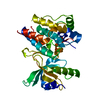
| ||||||||
|---|---|---|---|---|---|---|---|---|---|
| 1 |
| ||||||||
| Unit cell |
|
-Components
| #1: Protein | Mass: 38754.656 Da / Num. of mol.: 1 / Fragment: cytoplasmic region (residues 526-868) Source method: isolated from a genetically manipulated source Source: (gene. exp.)   Spodoptera frugiperda (fall armyworm) / References: UniProt: Q62838, EC: 2.7.1.112 Spodoptera frugiperda (fall armyworm) / References: UniProt: Q62838, EC: 2.7.1.112 |
|---|---|
| #2: Water | ChemComp-HOH /  Mass: 18.015 Da / Num. of mol.: 114 / Source method: isolated from a natural source / Formula: H2O Mass: 18.015 Da / Num. of mol.: 114 / Source method: isolated from a natural source / Formula: H2O |
-Experimental details
-Experiment
| Experiment | Method: X-RAY DIFFRACTION / Number of used crystals: 1 |
|---|
- Sample preparation
| Crystal | Density Matthews: 2.72 Å3/Da / Density % sol: 54.7 % | ||||||||||||||||||||||||||||||||||||||||||
|---|---|---|---|---|---|---|---|---|---|---|---|---|---|---|---|---|---|---|---|---|---|---|---|---|---|---|---|---|---|---|---|---|---|---|---|---|---|---|---|---|---|---|---|
| Crystal grow | Temperature: 277 K / Method: vapor diffusion, hanging drop / pH: 7 Details: ammonium sulfate, HEPES, glycerol, TCEP, pH 7.0, VAPOR DIFFUSION, HANGING DROP, temperature 277K | ||||||||||||||||||||||||||||||||||||||||||
| Crystal | *PLUS Density % sol: 58 % | ||||||||||||||||||||||||||||||||||||||||||
| Crystal grow | *PLUS Temperature: 20 ℃ | ||||||||||||||||||||||||||||||||||||||||||
| Components of the solutions | *PLUS
|
-Data collection
| Diffraction | Mean temperature: 100 K |
|---|---|
| Diffraction source | Source: SYNCHROTRON / Site: NSLS  / Beamline: X12C / Wavelength: 0.979 Å / Beamline: X12C / Wavelength: 0.979 Å |
| Detector | Type: CUSTOM-MADE / Detector: CCD / Date: Apr 29, 2001 |
| Radiation | Monochromator: silicon crystal / Protocol: SINGLE WAVELENGTH / Monochromatic (M) / Laue (L): M / Scattering type: x-ray |
| Radiation wavelength | Wavelength: 0.979 Å / Relative weight: 1 |
| Reflection | Resolution: 2.05→28 Å / Num. all: 28438 / Num. obs: 25737 / % possible obs: 99.7 % / Observed criterion σ(F): 1 / Observed criterion σ(I): 1 / Biso Wilson estimate: 14.1 Å2 |
| Reflection shell | Resolution: 2.05→2.1 Å / % possible all: 99 |
| Reflection | *PLUS Lowest resolution: 25 Å / Num. obs: 28438 / Num. measured all: 216370 / Rmerge(I) obs: 0.056 |
| Reflection shell | *PLUS % possible obs: 99.1 % / Rmerge(I) obs: 0.319 |
- Processing
| Software |
| ||||||||||||||||||||||||||||||||||||||||||||||||||||||||||||||||||||||||||||||||
|---|---|---|---|---|---|---|---|---|---|---|---|---|---|---|---|---|---|---|---|---|---|---|---|---|---|---|---|---|---|---|---|---|---|---|---|---|---|---|---|---|---|---|---|---|---|---|---|---|---|---|---|---|---|---|---|---|---|---|---|---|---|---|---|---|---|---|---|---|---|---|---|---|---|---|---|---|---|---|---|---|---|
| Refinement | Method to determine structure: MOLECULAR REPLACEMENT Starting model: PDB ID 1IRK Resolution: 2.05→26.31 Å / Rfactor Rfree error: 0.007 / Isotropic thermal model: RESTRAINED / Cross valid method: THROUGHOUT / σ(F): 2 / Stereochemistry target values: Engh & Huber
| ||||||||||||||||||||||||||||||||||||||||||||||||||||||||||||||||||||||||||||||||
| Solvent computation | Solvent model: FLAT MODEL / Bsol: 42.431 Å2 / ksol: 0.359255 e/Å3 | ||||||||||||||||||||||||||||||||||||||||||||||||||||||||||||||||||||||||||||||||
| Displacement parameters | Biso mean: 30.3 Å2
| ||||||||||||||||||||||||||||||||||||||||||||||||||||||||||||||||||||||||||||||||
| Refine analyze | Luzzati coordinate error free: 0.3 Å / Luzzati sigma a free: 0.27 Å | ||||||||||||||||||||||||||||||||||||||||||||||||||||||||||||||||||||||||||||||||
| Refinement step | Cycle: LAST / Resolution: 2.05→26.31 Å
| ||||||||||||||||||||||||||||||||||||||||||||||||||||||||||||||||||||||||||||||||
| Refine LS restraints |
| ||||||||||||||||||||||||||||||||||||||||||||||||||||||||||||||||||||||||||||||||
| LS refinement shell | Resolution: 2.05→2.18 Å / Rfactor Rfree error: 0.022 / Total num. of bins used: 6
| ||||||||||||||||||||||||||||||||||||||||||||||||||||||||||||||||||||||||||||||||
| Xplor file |
| ||||||||||||||||||||||||||||||||||||||||||||||||||||||||||||||||||||||||||||||||
| Refinement | *PLUS Lowest resolution: 25 Å / Num. reflection obs: 25737 / % reflection Rfree: 5 % / Rfactor obs: 0.231 / Rfactor Rfree: 0.252 / Rfactor Rwork: 0.234 | ||||||||||||||||||||||||||||||||||||||||||||||||||||||||||||||||||||||||||||||||
| Solvent computation | *PLUS | ||||||||||||||||||||||||||||||||||||||||||||||||||||||||||||||||||||||||||||||||
| Displacement parameters | *PLUS | ||||||||||||||||||||||||||||||||||||||||||||||||||||||||||||||||||||||||||||||||
| Refine LS restraints | *PLUS
| ||||||||||||||||||||||||||||||||||||||||||||||||||||||||||||||||||||||||||||||||
| LS refinement shell | *PLUS Lowest resolution: 2.1 Å / Rfactor Rfree: 0.313 / Rfactor Rwork: 0.27 |
