- PDB-1kmo: Crystal structure of the Outer Membrane Transporter FecA -
+
Open data
ID or keywords:
Loading...
-
Basic information
Entry
Database: PDB / ID: 1kmo
Title
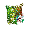
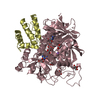
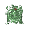
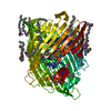
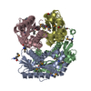
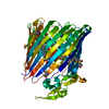
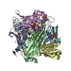
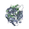
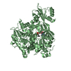
Crystal structure of the Outer Membrane Transporter FecA
Components
Iron(III) dicitrate transport protein fecA
Keywords
MEMBRANE PROTEIN / Iron transporter / TonB-dependent receptor / Siderophore
Function / homology
Function and homology information
response to iron ion starvation / siderophore-iron transmembrane transporter activity / siderophore-dependent iron import into cell / signal transduction involved in regulation of gene expression / transmembrane transporter complex / cell outer membrane / signaling receptor activity / iron ion transport / intracellular iron ion homeostasis / regulation of DNA-templated transcription / membrane Similarity search - Function
: / Secretin and TonB N terminus short domain / Secretin/TonB, short N-terminal domain / Secretin and TonB N terminus short domain / TonB-dependent receptor (TBDR) proteins signature 1. / TonB-dependent receptor, beta-barrel domain / TonB-dependent receptor, plug domain / Maltoporin; Chain A / TonB box, conserved site / Ferric Hydroxamate Uptake Protein; Chain A, domain 1 ...: / Secretin and TonB N terminus short domain / Secretin/TonB, short N-terminal domain / Secretin and TonB N terminus short domain / TonB-dependent receptor (TBDR) proteins signature 1. / TonB-dependent receptor, beta-barrel domain / TonB-dependent receptor, plug domain / Maltoporin; Chain A / TonB box, conserved site / Ferric Hydroxamate Uptake Protein; Chain A, domain 1 / TonB-dependent siderophore receptor / TonB-dependent receptor, conserved site / TonB-dependent receptor (TBDR) proteins signature 2. / TonB-dependent receptor (TBDR) proteins profile. / Vitamin B12 transporter BtuB-like / TonB-dependent receptor-like, beta-barrel / TonB dependent receptor-like, beta-barrel / TonB-dependent receptor, plug domain superfamily / TonB-dependent receptor, plug domain / TonB-dependent receptor-like, beta-barrel domain superfamily / TonB-dependent Receptor Plug Domain / Beta Complex / Beta Barrel / Mainly Beta Similarity search - Domain/homology
In the structure databanks used in Yorodumi, some data are registered as the other names, "COVID-19 virus" and "2019-nCoV". Here are the details of the virus and the list of structure data.
Jan 31, 2019. EMDB accession codes are about to change! (news from PDBe EMDB page)
EMDB accession codes are about to change! (news from PDBe EMDB page)
The allocation of 4 digits for EMDB accession codes will soon come to an end. Whilst these codes will remain in use, new EMDB accession codes will include an additional digit and will expand incrementally as the available range of codes is exhausted. The current 4-digit format prefixed with “EMD-” (i.e. EMD-XXXX) will advance to a 5-digit format (i.e. EMD-XXXXX), and so on. It is currently estimated that the 4-digit codes will be depleted around Spring 2019, at which point the 5-digit format will come into force.
The EM Navigator/Yorodumi systems omit the EMD- prefix.
Related info.:Q: What is EMD? / ID/Accession-code notation in Yorodumi/EM Navigator
Yorodumi is a browser for structure data from EMDB, PDB, SASBDB, etc.
This page is also the successor to EM Navigator detail page, and also detail information page/front-end page for Omokage search.
The word "yorodu" (or yorozu) is an old Japanese word meaning "ten thousand". "mi" (miru) is to see.
Related info.:EMDB / PDB / SASBDB / Comparison of 3 databanks / Yorodumi Search / Aug 31, 2016. New EM Navigator & Yorodumi / Yorodumi Papers / Jmol/JSmol / Function and homology information / Changes in new EM Navigator and Yorodumi
 Movie
Movie Controller


 Open data
 Basic information
 Components
 Keywords
 Function and homology information
 X-RAY DIFFRACTION /
  Authors
 Citation
 Structure visualization
 Downloads & links
 Other downloads

 PDBj
PDBj

 Assembly
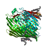

 Mass: 229.402 Da / Num. of mol.: 17 / Source method: obtained synthetically / Formula: C14H31NO / Comment: LDAO, detergent*YM
 Mass: 148.200 Da / Num. of mol.: 2 / Source method: obtained synthetically / Formula: C7H16O3
 Mass: 18.015 Da / Num. of mol.: 386 / Source method: isolated from a natural source / Formula: H2O
 Sample preparation
 / Beamline: X12B / Wavelength: 0.978 Å
 Processing