 Movie
Movie Controller


+ Open data

- Basic information
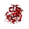
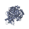
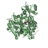
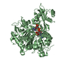
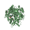
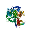
| Entry | Database: PDB / ID: 2p20 | ||||||
|---|---|---|---|---|---|---|---|
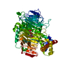
| Title | Acetyl-CoA Synthetase, R584A mutation | ||||||
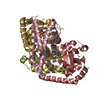
 Components Components | Acetyl-coenzyme A synthetase | ||||||
 Keywords Keywords | LIGASE / Adenylate-forming enzymes / Domain Alternation / Acyl-CoA ligase | ||||||
| Function / homology |  Function and homology information Function and homology informationacetate-CoA ligase / acetyl-CoA synthetase activity / : / acetyl-CoA biosynthetic process / AMP binding / chemotaxis / ATP binding / metal ion binding / cytosol Similarity search - Function | ||||||
| Biological species |  Salmonella typhimurium (bacteria) Salmonella typhimurium (bacteria) | ||||||
| Method |  X-RAY DIFFRACTION / SYNCHROTRON / MOLECULAR REPLACEMENT / Resolution: 2.22 Å X-RAY DIFFRACTION / SYNCHROTRON / MOLECULAR REPLACEMENT / Resolution: 2.22 Å | ||||||
 Authors Authors | Reger, A.S. / Gulick, A.M. | ||||||
 Citation Citation | Journal: Biochemistry / Year: 2007 Title: Biochemical and Crystallographic Analysis of Substrate Binding and Conformational Changes in Acetyl-CoA Synthetase. Authors: Reger, A.S. / Carney, J.M. / Gulick, A.M. | ||||||
| History |
|
- Structure visualization
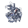
| Structure viewer | Molecule: MolmilJmol/JSmol |
|---|
- Downloads & links
-Download
| PDBx/mmCIF format | 2p20.cif.gz | 265.7 KB | Display | PDBx/mmCIF format |
|---|---|---|---|---|
| PDB format | pdb2p20.ent.gz | 211.4 KB | Display | PDB format |
| PDBx/mmJSON format | 2p20.json.gz | Tree view | PDBx/mmJSON format | |
| Others |  Other downloads Other downloads |
-Validation report
| Arichive directory | https://data.pdbj.org/pub/pdb/validation_reports/p2/2p20ftp://data.pdbj.org/pub/pdb/validation_reports/p2/2p20 | HTTPS FTP |
|---|
-Related structure data
| Related structure data |  2p2bC  2p2fC  2p2jC  2p2mC  2p2qC 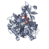 1pg4S S: Starting model for refinement C: citing same article ( |
|---|---|
| Similar structure data |
-Links
 PDBj
PDBj
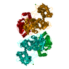
- Assembly
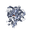
| Deposited unit | 
| ||||||||
|---|---|---|---|---|---|---|---|---|---|
| 1 | 
| ||||||||
| 2 | 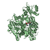
| ||||||||
| Unit cell |
|
-Components
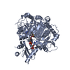
| #1: Protein | Mass: 72150.359 Da / Num. of mol.: 2 / Mutation: R584A Source method: isolated from a genetically manipulated source Source: (gene. exp.) Salmonella typhimurium (bacteria) / Strain: Typhi / Gene: acs / Plasmid: pTYB1 / Species (production host): Escherichia coli / Production host: #2: Chemical |   Mass: 389.301 Da / Num. of mol.: 2 / Source method: obtained synthetically / Formula: C13H20N5O7P / Details: according to Biochemistry, 41, 2379. Mass: 389.301 Da / Num. of mol.: 2 / Source method: obtained synthetically / Formula: C13H20N5O7P / Details: according to Biochemistry, 41, 2379.#3: Water | ChemComp-HOH / |  Mass: 18.015 Da / Num. of mol.: 600 / Source method: isolated from a natural source / Formula: H2O Mass: 18.015 Da / Num. of mol.: 600 / Source method: isolated from a natural source / Formula: H2O |
|---|
-Experimental details
-Experiment
| Experiment | Method: X-RAY DIFFRACTION / Number of used crystals: 1 |
|---|
- Sample preparation
| Crystal | Density Matthews: 2.5 Å3/Da / Density % sol: 50.78 % |
|---|---|
| Crystal grow | Temperature: 287 K / Method: vapor diffusion, hanging drop / pH: 6.5 Details: 6-15% PEG 8000, 9-13% ethylene glycol, 50 mM BTP, 1 mM CoA, 1 mM Propyl-AMP, 1 mM TCEP, pH 6.5, VAPOR DIFFUSION, HANGING DROP, temperature 287K |
-Data collection
| Diffraction | Mean temperature: 113 K |
|---|---|
| Diffraction source | Source: SYNCHROTRON / Site: CHESS  / Beamline: F2 / Wavelength: 0.9793 Å / Beamline: F2 / Wavelength: 0.9793 Å |
| Detector | Type: ADSC QUANTUM 210 / Detector: CCD / Date: Jul 31, 2005 |
| Radiation | Protocol: SINGLE WAVELENGTH / Monochromatic (M) / Laue (L): M / Scattering type: x-ray |
| Radiation wavelength | Wavelength: 0.9793 Å / Relative weight: 1 |
| Reflection | Resolution: 2.22→66.227 Å / Num. obs: 67992 / % possible obs: 92 % / Redundancy: 4.6 % / Biso Wilson estimate: 26.3 Å2 / Rmerge(I) obs: 0.062 / Rsym value: 0.062 / Net I/σ(I): 8.4 |
| Reflection shell | Resolution: 2.22→2.32 Å / Redundancy: 3 % / Rmerge(I) obs: 0.314 / Mean I/σ(I) obs: 0.9 / Num. measured all: 19336 / Num. unique all: 6481 / Rsym value: 0.314 / % possible all: 61.6 |
- Processing
| Software |
| |||||||||||||||||||||||||||||||||||||||||||||||||||||||||||||||||||||||||||||||||||||||||||||||||||||||||||||||||||||||||||||
|---|---|---|---|---|---|---|---|---|---|---|---|---|---|---|---|---|---|---|---|---|---|---|---|---|---|---|---|---|---|---|---|---|---|---|---|---|---|---|---|---|---|---|---|---|---|---|---|---|---|---|---|---|---|---|---|---|---|---|---|---|---|---|---|---|---|---|---|---|---|---|---|---|---|---|---|---|---|---|---|---|---|---|---|---|---|---|---|---|---|---|---|---|---|---|---|---|---|---|---|---|---|---|---|---|---|---|---|---|---|---|---|---|---|---|---|---|---|---|---|---|---|---|---|---|---|---|
| Refinement | Method to determine structure: MOLECULAR REPLACEMENT Starting model: pdb entry 1PG4 Resolution: 2.22→30 Å / Cor.coef. Fo:Fc: 0.943 / Cor.coef. Fo:Fc free: 0.905 / SU B: 11.1 / SU ML: 0.147 / TLS residual ADP flag: LIKELY RESIDUAL / Cross valid method: THROUGHOUT / σ(F): 0 / ESU R: 0.287 / ESU R Free: 0.217 / Stereochemistry target values: MAXIMUM LIKELIHOOD / Details: HYDROGENS HAVE BEEN ADDED IN THE RIDING POSITIONS
| |||||||||||||||||||||||||||||||||||||||||||||||||||||||||||||||||||||||||||||||||||||||||||||||||||||||||||||||||||||||||||||
| Solvent computation | Ion probe radii: 0.8 Å / Shrinkage radii: 0.8 Å / VDW probe radii: 1.2 Å / Solvent model: BABINET MODEL WITH MASK | |||||||||||||||||||||||||||||||||||||||||||||||||||||||||||||||||||||||||||||||||||||||||||||||||||||||||||||||||||||||||||||
| Displacement parameters | Biso mean: 18.929 Å2
| |||||||||||||||||||||||||||||||||||||||||||||||||||||||||||||||||||||||||||||||||||||||||||||||||||||||||||||||||||||||||||||
| Refinement step | Cycle: LAST / Resolution: 2.22→30 Å
| |||||||||||||||||||||||||||||||||||||||||||||||||||||||||||||||||||||||||||||||||||||||||||||||||||||||||||||||||||||||||||||
| Refine LS restraints |
| |||||||||||||||||||||||||||||||||||||||||||||||||||||||||||||||||||||||||||||||||||||||||||||||||||||||||||||||||||||||||||||
| LS refinement shell | Resolution: 2.22→2.272 Å / Total num. of bins used: 20
| |||||||||||||||||||||||||||||||||||||||||||||||||||||||||||||||||||||||||||||||||||||||||||||||||||||||||||||||||||||||||||||
| Refinement TLS params. | Method: refined / Refine-ID: X-RAY DIFFRACTION
| |||||||||||||||||||||||||||||||||||||||||||||||||||||||||||||||||||||||||||||||||||||||||||||||||||||||||||||||||||||||||||||
| Refinement TLS group | Refine-ID: X-RAY DIFFRACTION / Selection: ALL
|
