 Movie
Movie Controller

[English] 日本語
 Yorodumi
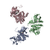
- PDB-1ry2: Crystal structure of yeast acetyl-coenzyme A synthetase in comple... -
+ Open data

- Basic information
| Entry | Database: PDB / ID: 1ry2 | ||||||
|---|---|---|---|---|---|---|---|
| Title | Crystal structure of yeast acetyl-coenzyme A synthetase in complex with AMP | ||||||
 Components Components | acetyl-coenzyme A synthetase 1 | ||||||
 Keywords Keywords | LIGASE / amp forming / related to firefly luciferase | ||||||
| Function / homology |  Function and homology information Function and homology informationpyruvate fermentation to acetate / Ethanol oxidation / acid-ammonia (or amide) ligase activity / : / acetate-CoA ligase / acetyl-CoA synthetase activity / : / acetyl-CoA biosynthetic process / AMP binding / mitochondrion ...pyruvate fermentation to acetate / Ethanol oxidation / acid-ammonia (or amide) ligase activity / : / acetate-CoA ligase / acetyl-CoA synthetase activity / : / acetyl-CoA biosynthetic process / AMP binding / mitochondrion / ATP binding / nucleus / cytosol Similarity search - Function | ||||||
| Biological species |  | ||||||
| Method |  X-RAY DIFFRACTION / SYNCHROTRON / MOLECULAR REPLACEMENT / Resolution: 2.3 Å X-RAY DIFFRACTION / SYNCHROTRON / MOLECULAR REPLACEMENT / Resolution: 2.3 Å | ||||||
 Authors Authors | Jogl, G. / Tong, L. | ||||||
 Citation Citation | Journal: Biochemistry / Year: 2004 Title: Crystal structure of yeast acetyl-coenzyme A synthetase in complex with AMP Authors: Jogl, G. / Tong, L. | ||||||
| History |
|
- Structure visualization
| Structure viewer | Molecule: MolmilJmol/JSmol |
|---|
- Downloads & links
-Download
| PDBx/mmCIF format | 1ry2.cif.gz | 134.5 KB | Display | PDBx/mmCIF format |
|---|---|---|---|---|
| PDB format | pdb1ry2.ent.gz | 102.8 KB | Display | PDB format |
| PDBx/mmJSON format | 1ry2.json.gz | Tree view | PDBx/mmJSON format | |
| Others |  Other downloads Other downloads |
-Validation report
| Arichive directory | https://data.pdbj.org/pub/pdb/validation_reports/ry/1ry2ftp://data.pdbj.org/pub/pdb/validation_reports/ry/1ry2 | HTTPS FTP |
|---|
-Related structure data
| Related structure data | 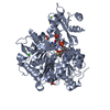 1pg4S S: Starting model for refinement |
|---|---|
| Similar structure data |


-Links
 PDBj
PDBj


- Assembly
| Deposited unit | 
| ||||||||||
|---|---|---|---|---|---|---|---|---|---|---|---|
| 1 | 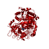
| ||||||||||
| Unit cell |
| ||||||||||
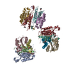
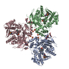
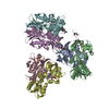
| Details | biological assembly is a trimer generated by space group symm -y, x-y, z -x+y, -x, z |
-Components
| #1: Protein | Mass: 73603.195 Da / Num. of mol.: 1 Source method: isolated from a genetically manipulated source Source: (gene. exp.) Plasmid: pET28a / Production host:  |
|---|---|
| #2: Chemical | ChemComp-AMP /   Mass: 347.221 Da / Num. of mol.: 1 / Source method: obtained synthetically / Formula: C10H14N5O7P / Comment: AMP*YM Mass: 347.221 Da / Num. of mol.: 1 / Source method: obtained synthetically / Formula: C10H14N5O7P / Comment: AMP*YM |
-Experimental details
-Experiment
| Experiment | Method: X-RAY DIFFRACTION / Number of used crystals: 1 |
|---|
- Sample preparation
| Crystal | Density Matthews: 2.67 Å3/Da / Density % sol: 53.6 % | ||||||||||||||||||||||||||||||
|---|---|---|---|---|---|---|---|---|---|---|---|---|---|---|---|---|---|---|---|---|---|---|---|---|---|---|---|---|---|---|---|
| Crystal grow | Temperature: 277 K / Method: vapor diffusion, sitting drop / pH: 7.5 Details: succinic acid, tris, pH 7.5, VAPOR DIFFUSION, SITTING DROP, temperature 271K | ||||||||||||||||||||||||||||||
| Crystal grow | *PLUS Temperature: 4 ℃ / Method: vapor diffusion, sitting drop | ||||||||||||||||||||||||||||||
| Components of the solutions | *PLUS
|
-Data collection
| Diffraction | Mean temperature: 100 K |
|---|---|
| Diffraction source | Source: SYNCHROTRON / Site: NSLS  / Beamline: X4A / Wavelength: 0.9791 Å / Beamline: X4A / Wavelength: 0.9791 Å |
| Detector | Type: ADSC QUANTUM 4 / Detector: CCD / Date: Aug 21, 2003 |
| Radiation | Monochromator: Si 111 CHANNEL / Protocol: SINGLE WAVELENGTH / Monochromatic (M) / Laue (L): M / Scattering type: x-ray |
| Radiation wavelength | Wavelength: 0.9791 Å / Relative weight: 1 |
| Reflection | Resolution: 2.3→30 Å / Num. all: 27899 / Num. obs: 27899 / % possible obs: 89.3 % / Observed criterion σ(F): 0 / Observed criterion σ(I): 0 / Biso Wilson estimate: 26.9 Å2 |
| Reflection shell | Resolution: 2.3→2.44 Å / % possible all: 79.1 |
| Reflection | *PLUS Highest resolution: 2.3 Å / % possible obs: 89 % / Num. measured all: 85039 / Rmerge(I) obs: 0.066 |
| Reflection shell | *PLUS % possible obs: 79 % / Rmerge(I) obs: 0.24 |
- Processing
| Software |
| ||||||||||||||||||||||||||||||||||||
|---|---|---|---|---|---|---|---|---|---|---|---|---|---|---|---|---|---|---|---|---|---|---|---|---|---|---|---|---|---|---|---|---|---|---|---|---|---|
| Refinement | Method to determine structure: MOLECULAR REPLACEMENT Starting model: pdb entry 1pg4 Resolution: 2.3→29.54 Å / Rfactor Rfree error: 0.006 / Data cutoff high absF: 316294.72 / Data cutoff low absF: 0 / Isotropic thermal model: RESTRAINED / Cross valid method: THROUGHOUT / σ(F): 0 / Stereochemistry target values: Engh & Huber / Details: BULK SOLVENT MODEL USED
| ||||||||||||||||||||||||||||||||||||
| Solvent computation | Solvent model: FLAT MODEL / Bsol: 43.4283 Å2 / ksol: 0.306315 e/Å3 | ||||||||||||||||||||||||||||||||||||
| Displacement parameters | Biso mean: 40.6 Å2
| ||||||||||||||||||||||||||||||||||||
| Refine analyze |
| ||||||||||||||||||||||||||||||||||||
| Refinement step | Cycle: LAST / Resolution: 2.3→29.54 Å
| ||||||||||||||||||||||||||||||||||||
| Refine LS restraints |
| ||||||||||||||||||||||||||||||||||||
| LS refinement shell | Resolution: 2.3→2.44 Å / Rfactor Rfree error: 0.019 / Total num. of bins used: 6
| ||||||||||||||||||||||||||||||||||||
| Xplor file |
| ||||||||||||||||||||||||||||||||||||
| Refinement | *PLUS Highest resolution: 2.3 Å / Lowest resolution: 30 Å | ||||||||||||||||||||||||||||||||||||
| Solvent computation | *PLUS | ||||||||||||||||||||||||||||||||||||
| Displacement parameters | *PLUS | ||||||||||||||||||||||||||||||||||||
| Refine LS restraints | *PLUS
|
