 Movie
Movie Controller

[English] 日本語
 Yorodumi
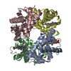
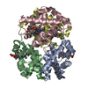
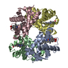
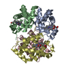
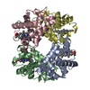
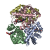
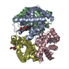
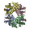
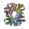
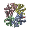
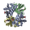
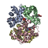
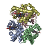
- PDB-1kd2: Crystal Structure of Human Deoxyhemoglobin in Absence of Any Anions -
+ Open data

- Basic information
| Entry | Database: PDB / ID: 1kd2 | ||||||
|---|---|---|---|---|---|---|---|
| Title | Crystal Structure of Human Deoxyhemoglobin in Absence of Any Anions | ||||||
 Components Components |
| ||||||
 Keywords Keywords | OXYGEN STORAGE/TRANSPORT / HUMAN DEOXY HEMOGLOBIN / HIGH O2 AFFINITY T STATE / CHLORIDE BINDING SITES / ALLOSTERIC ANION BINDING SITES / HYDRATION / OXYGEN STORAGE-TRANSPORT COMPLEX | ||||||
| Function / homology |  Function and homology information Function and homology informationnitric oxide transport / hemoglobin alpha binding / haptoglobin-hemoglobin complex /  organic acid binding / cellular oxidant detoxification / hemoglobin binding / renal absorption / hemoglobin complex / oxygen transport / Scavenging of heme from plasma ...nitric oxide transport / hemoglobin alpha binding / haptoglobin-hemoglobin complex / organic acid binding / cellular oxidant detoxification / hemoglobin binding / renal absorption / hemoglobin complex / oxygen transport / Scavenging of heme from plasma / endocytic vesicle lumen / blood vessel diameter maintenance / hydrogen peroxide catabolic process / oxygen carrier activity / Late endosomal microautophagy / Heme signaling / carbon dioxide transport / response to hydrogen peroxide / Erythrocytes take up oxygen and release carbon dioxide / Erythrocytes take up carbon dioxide and release oxygen / Cytoprotection by HMOX1 / platelet aggregation / oxygen binding / regulation of blood pressure / Chaperone Mediated Autophagy / positive regulation of nitric oxide biosynthetic process / tertiary granule lumen / Factors involved in megakaryocyte development and platelet production / blood microparticle / ficolin-1-rich granule lumen / iron ion binding / heme binding / Neutrophil degranulation / extracellular space / extracellular exosome / extracellular region / membrane / metal ion binding / cytosol organic acid binding / cellular oxidant detoxification / hemoglobin binding / renal absorption / hemoglobin complex / oxygen transport / Scavenging of heme from plasma ...nitric oxide transport / hemoglobin alpha binding / haptoglobin-hemoglobin complex / organic acid binding / cellular oxidant detoxification / hemoglobin binding / renal absorption / hemoglobin complex / oxygen transport / Scavenging of heme from plasma / endocytic vesicle lumen / blood vessel diameter maintenance / hydrogen peroxide catabolic process / oxygen carrier activity / Late endosomal microautophagy / Heme signaling / carbon dioxide transport / response to hydrogen peroxide / Erythrocytes take up oxygen and release carbon dioxide / Erythrocytes take up carbon dioxide and release oxygen / Cytoprotection by HMOX1 / platelet aggregation / oxygen binding / regulation of blood pressure / Chaperone Mediated Autophagy / positive regulation of nitric oxide biosynthetic process / tertiary granule lumen / Factors involved in megakaryocyte development and platelet production / blood microparticle / ficolin-1-rich granule lumen / iron ion binding / heme binding / Neutrophil degranulation / extracellular space / extracellular exosome / extracellular region / membrane / metal ion binding / cytosolSimilarity search - Function | ||||||
| Biological species |  Homo sapiens (human) Homo sapiens (human) | ||||||
| Method | X-RAY DIFFRACTION / SYNCHROTRON / MOLECULAR REPLACEMENT / Resolution: 1.87 Å | ||||||
 Authors Authors | Colombo, M.F. / Seixas, F.A.V. | ||||||
 Citation Citation | Journal: To be Published Title: The X-Ray Structure of Iso-Ionic Deoxy-Hb Crystal: The High Affinity T-state of Human Hb and the Mechanism of Chloride Regulation of Hb Cooperative Oxygen Binding. Authors: Seixas, F.A.V. / Azevedo Jr, W.F. / Colombo, M.F. #1: Journal: Acta Crystallogr.,Sect.D / Year: 1999Title: Crystallization and X-ray Diffraction Data Analysis of Human Deoxyhaemoglobin A(0) Fully stripped of Any Anions Authors: Seixas, F.A.V. / Azevedo Jr, W.F. / Colombo, M.F. | ||||||
| History |
|
- Structure visualization
| Structure viewer | Molecule: MolmilJmol/JSmol |
|---|
- Downloads & links
-Download
| PDBx/mmCIF format | 1kd2.cif.gz | 123.5 KB | Display | PDBx/mmCIF format |
|---|---|---|---|---|
| PDB format | pdb1kd2.ent.gz | 98 KB | Display | PDB format |
| PDBx/mmJSON format | 1kd2.json.gz | Tree view | PDBx/mmJSON format | |
| Others |  Other downloads Other downloads |
-Validation report
| Arichive directory | https://data.pdbj.org/pub/pdb/validation_reports/kd/1kd2ftp://data.pdbj.org/pub/pdb/validation_reports/kd/1kd2 | HTTPS FTP |
|---|
-Related structure data
| Related structure data |  2hhbS S: Starting model for refinement |
|---|---|
| Similar structure data |


-Links
 PDBj
PDBj


- Assembly
| Deposited unit | 
| ||||||||
|---|---|---|---|---|---|---|---|---|---|
| 1 |
| ||||||||
| Unit cell |
| ||||||||
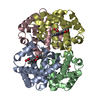
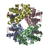
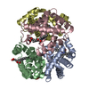
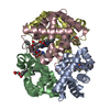
| Details | THE CRYSTALLOGRAPHIC ASYMMETRIC UNIT CONTAINS TWO ALPHA AND TWO BETA CHAINS. |
-Components
| #1: Protein | Mass: 15150.353 Da / Num. of mol.: 2 / Source method: isolated from a natural source / Source: (natural) Homo sapiens (human) / Tissue: BLOOD / References: UniProt: P69905#2: Protein | Mass: 15890.198 Da / Num. of mol.: 2 / Source method: isolated from a natural source / Source: (natural) Homo sapiens (human) / Tissue: BLOOD / References: UniProt: P68871#3: Chemical | ChemComp-HEM / Heme B  Mass: 616.487 Da / Num. of mol.: 4 / Source method: obtained synthetically / Formula: C34H32FeN4O4 Mass: 616.487 Da / Num. of mol.: 4 / Source method: obtained synthetically / Formula: C34H32FeN4O4#4: Water | ChemComp-HOH / | Water Mass: 18.015 Da / Num. of mol.: 161 / Source method: isolated from a natural source / Formula: H2O Mass: 18.015 Da / Num. of mol.: 161 / Source method: isolated from a natural source / Formula: H2O |
|---|
-Experimental details
-Experiment
| Experiment | Method: X-RAY DIFFRACTION / Number of used crystals: 1 |
|---|
- Sample preparation
| Crystal | Density Matthews: 2.48 Å3/Da / Density % sol: 50.4 % |
|---|---|
| Crystal grow | Temperature: 298 K / Method: batch method / pH: 7 Details: DEIONIZED PEG 3350 12.5%, pH 7.0, BATCH METHOD, temperature 298K |
-Data collection
| Diffraction | Mean temperature: 299 K |
|---|---|
| Diffraction source | Source: SYNCHROTRON / Site: LNLS  / Beamline: D03B-MX1 / Wavelength: 1.38 Å / Beamline: D03B-MX1 / Wavelength: 1.38 Å |
| Detector | Type: MARRESEARCH / Detector: IMAGE PLATE / Date: Aug 26, 1998 |
| Radiation | Monochromator: SILICON CRYSTAL WITH ASYMMETRIC CUT. ASYMMETRIC ANGLE: 7.25 DEGREES, CONDENSATE MODE Protocol: SINGLE WAVELENGTH / Monochromatic (M) / Laue (L): M / Scattering type: x-ray |
| Radiation wavelength | Wavelength: 1.38 Å / Relative weight: 1 |
| Reflection | Resolution: 1.87→69.007 Å / Num. all: 51413 / Num. obs: 51030 / % possible obs: 95.6 % / Observed criterion σ(F): 2 / Observed criterion σ(I): 2.8 / Rsym value: 0.062 |
| Reflection shell | Resolution: 1.87→1.91 Å / Num. unique all: 3240 / Rsym value: 0.348 / % possible all: 92.6 |
- Processing
| Software |
| |||||||||||||||||||||
|---|---|---|---|---|---|---|---|---|---|---|---|---|---|---|---|---|---|---|---|---|---|---|
| Refinement | Method to determine structure: MOLECULAR REPLACEMENT Starting model: PDB ENTRY 2HHB Resolution: 1.87→10 Å / Isotropic thermal model: overall temperature factors / σ(F): 2 / Stereochemistry target values: Engh & Huber
| |||||||||||||||||||||
| Displacement parameters | Biso mean: 23.062 Å2 | |||||||||||||||||||||
| Refine analyze | Luzzati d res low obs: 5 Å / Luzzati sigma a obs: 0.28 Å | |||||||||||||||||||||
| Refinement step | Cycle: LAST / Resolution: 1.87→10 Å
| |||||||||||||||||||||
| Refine LS restraints |
| |||||||||||||||||||||
| LS refinement shell | Resolution: 1.87→1.91 Å
|
