Mass: 15.999 Da / Num. of mol.: 2 / Source method: obtained synthetically / Formula: O
Nonpolymer details
RESIDUES 339 - 346, 475 - 482, 885 - 891 AND 1041 - 1046 AND THE CARBOHYDRATE SIDE CHAINS ATTACHED ...RESIDUES 339 - 346, 475 - 482, 885 - 891 AND 1041 - 1046 AND THE CARBOHYDRATE SIDE CHAINS ATTACHED TO ASN 339 AND ASN 378 ARE MISSING DUE TO BREAKS IN THE ELECTRON DENSITY. ONLY THE FIRST NAG RESIDUE HAS BEEN PLACED FOR ASN 119 AND AND ASN 743.
-
Experimental details
-
Experiment
Experiment
Method: X-RAY DIFFRACTION / Number of used crystals: 6
-
Sample preparation
Crystal
Density Matthews: 4.3 Å3/Da / Density % sol: 70 %
Crystal grow
pH: 5.65 / Details: pH 5.65
Crystal grow
*PLUS
Temperature: 277 K / pH: 5.45 / Method: vapor diffusion, sitting drop
In the structure databanks used in Yorodumi, some data are registered as the other names, "COVID-19 virus" and "2019-nCoV". Here are the details of the virus and the list of structure data.
Jan 31, 2019. EMDB accession codes are about to change! (news from PDBe EMDB page)
EMDB accession codes are about to change! (news from PDBe EMDB page)
The allocation of 4 digits for EMDB accession codes will soon come to an end. Whilst these codes will remain in use, new EMDB accession codes will include an additional digit and will expand incrementally as the available range of codes is exhausted. The current 4-digit format prefixed with “EMD-” (i.e. EMD-XXXX) will advance to a 5-digit format (i.e. EMD-XXXXX), and so on. It is currently estimated that the 4-digit codes will be depleted around Spring 2019, at which point the 5-digit format will come into force.
The EM Navigator/Yorodumi systems omit the EMD- prefix.
Related info.:Q: What is EMD? / ID/Accession-code notation in Yorodumi/EM Navigator
Yorodumi is a browser for structure data from EMDB, PDB, SASBDB, etc.
This page is also the successor to EM Navigator detail page, and also detail information page/front-end page for Omokage search.
The word "yorodu" (or yorozu) is an old Japanese word meaning "ten thousand". "mi" (miru) is to see.
Related info.:EMDB / PDB / SASBDB / Comparison of 3 databanks / Yorodumi Search / Aug 31, 2016. New EM Navigator & Yorodumi / Yorodumi Papers / Jmol/JSmol / Function and homology information / Changes in new EM Navigator and Yorodumi
 Movie
Movie Controller


 Open data
 Basic information
 Components
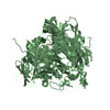
 Keywords
 Function and homology information
 Authors
 Citation
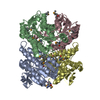
 Structure visualization
 Downloads & links
 Other downloads
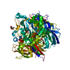


 PDBj
PDBj


 Assembly

 Type: D-saccharide, beta linking / Mass: 221.208 Da / Num. of mol.: 2
 Mass: 63.546 Da / Num. of mol.: 8 / Source method: obtained synthetically / Formula: Cu
 Mass: 15.999 Da / Num. of mol.: 2 / Source method: obtained synthetically / Formula: O
 Sample preparation
 / Beamline: PX9.6 / Wavelength: 0.87 / Wavelength: 0.87, 0.882
 Processing