 Movie
Movie Controller

[English] 日本語
 Yorodumi
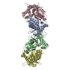
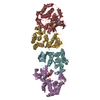
- PDB-1jx2: CRYSTAL STRUCTURE OF THE NUCLEOTIDE-FREE DYNAMIN A GTPASE DOMAIN,... -
+ Open data

- Basic information
| Entry | Database: PDB / ID: 1jx2 | ||||||
|---|---|---|---|---|---|---|---|
| Title | CRYSTAL STRUCTURE OF THE NUCLEOTIDE-FREE DYNAMIN A GTPASE DOMAIN, DETERMINED AS MYOSIN FUSION | ||||||
 Components Components | Myosin-2 heavy chain,Dynamin-A | ||||||
 Keywords Keywords | HYDROLASE / dynamin / GTPase / myosin / fusion-protein / Dictyostelium | ||||||
| Function / homology |  Function and homology information Function and homology informationprotein processing in phagocytic vesicle / Regulation of Apoptosis / Apoptotic execution phase / sorocarp morphogenesis / regulation of post-lysosomal vacuole size / phagosome acidification / ISG15 antiviral mechanism / protein localization to cleavage furrow / pinocytosis / uropod retraction ...protein processing in phagocytic vesicle / Regulation of Apoptosis / Apoptotic execution phase / sorocarp morphogenesis / regulation of post-lysosomal vacuole size / phagosome acidification / ISG15 antiviral mechanism / protein localization to cleavage furrow / pinocytosis / uropod retraction / cytoplasmic actin-based contraction involved in forward cell motility / phagocytic cup base / pathogen-containing vacuole / response to differentiation-inducing factor 1 / equatorial cell cortex / contractile actin filament bundle assembly / pseudopodium retraction / cell trailing edge / contractile vacuole organization / myosin filament assembly / aggregation involved in sorocarp development / culmination involved in sorocarp development / adenyl nucleotide binding / calcium-dependent ATPase activity / RHO GTPases activate PAKs / hypotonic response / actomyosin contractile ring / uropod / apical cortex / peroxisome fission / profilin binding / negative regulation of actin filament polymerization / actin-myosin filament sliding / detection of mechanical stimulus / substrate-dependent cell migration, cell extension / midbody abscission / bleb assembly / actomyosin / filopodium assembly / endosome organization / myosin filament / early phagosome / mitochondrial fission / myosin II complex / cortical actin cytoskeleton organization / microfilament motor activity / cortical actin cytoskeleton / nucleus organization / intracellular distribution of mitochondria / establishment or maintenance of cell polarity / pseudopodium / cleavage furrow / cytoskeletal motor activity / mitotic cytokinesis / response to cAMP / response to mechanical stimulus / phagocytosis / intercellular bridge / phagocytic vesicle / 14-3-3 protein binding / actin filament organization / mitochondrion organization / response to bacterium / cell motility / response to hydrogen peroxide / phospholipid binding / extracellular matrix / chemotaxis / actin filament binding / intracellular protein localization / regulation of cell shape / cytoplasmic vesicle / cell cortex / microtubule binding / microtubule / cytoskeleton / calmodulin binding / GTPase activity / GTP binding / ATP binding / identical protein binding / membrane / cytoplasm / cytosol Similarity search - Function | ||||||
| Biological species |  | ||||||
| Method |  X-RAY DIFFRACTION / SYNCHROTRON / MOLECULAR REPLACEMENT / Resolution: 2.3 Å X-RAY DIFFRACTION / SYNCHROTRON / MOLECULAR REPLACEMENT / Resolution: 2.3 Å | ||||||
 Authors Authors | Niemann, H.H. / Knetsch, M.L.W. / Scherer, A. / Manstein, D.J. / Kull, F.J. | ||||||
 Citation Citation | Journal: EMBO J. / Year: 2001 Title: Crystal structure of a dynamin GTPase domain in both nucleotide-free and GDP-bound forms. Authors: Niemann, H.H. / Knetsch, M.L. / Scherer, A. / Manstein, D.J. / Kull, F.J. | ||||||
| History |
| ||||||
| Remark 999 | SEQUENCE According to the author, Electron density map confirm residue 260 to be SER and residue ...SEQUENCE According to the author, Electron density map confirm residue 260 to be SER and residue 323 to be CYS. |
- Structure visualization
| Structure viewer | Molecule: MolmilJmol/JSmol |
|---|
- Downloads & links
-Download
| PDBx/mmCIF format | 1jx2.cif.gz | 232.9 KB | Display | PDBx/mmCIF format |
|---|---|---|---|---|
| PDB format | pdb1jx2.ent.gz | 180.2 KB | Display | PDB format |
| PDBx/mmJSON format | 1jx2.json.gz | Tree view | PDBx/mmJSON format | |
| Others |  Other downloads Other downloads |
-Validation report
| Arichive directory | https://data.pdbj.org/pub/pdb/validation_reports/jx/1jx2ftp://data.pdbj.org/pub/pdb/validation_reports/jx/1jx2 | HTTPS FTP |
|---|
-Related structure data
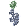
| Related structure data | 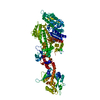 1jwyC 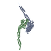 1g8xS C: citing same article ( S: Starting model for refinement |
|---|---|
| Similar structure data |


-Links
 PDBj
PDBj


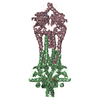
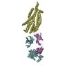
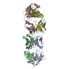
- Assembly
| Deposited unit | 
| ||||||||
|---|---|---|---|---|---|---|---|---|---|
| 1 |
| ||||||||
| Unit cell |
|
-Components
| #1: Protein | Mass: 124573.531 Da / Num. of mol.: 1 / Fragment: catalytic domain Source method: isolated from a genetically manipulated source Source: (gene. exp.) |
|---|---|
| #2: Sugar | ChemComp-BGC /   Type: D-saccharide, beta linking / Mass: 180.156 Da / Num. of mol.: 1 Type: D-saccharide, beta linking / Mass: 180.156 Da / Num. of mol.: 1Source method: isolated from a genetically manipulated source Formula: C6H12O6 |
| #3: Chemical | ChemComp-MG /   Mass: 24.305 Da / Num. of mol.: 1 / Source method: obtained synthetically / Formula: Mg Mass: 24.305 Da / Num. of mol.: 1 / Source method: obtained synthetically / Formula: Mg |
| #4: Chemical | ChemComp-ADP /   Mass: 427.201 Da / Num. of mol.: 1 / Source method: obtained synthetically / Formula: C10H15N5O10P2 / Comment: ADP, energy-carrying molecule*YM Mass: 427.201 Da / Num. of mol.: 1 / Source method: obtained synthetically / Formula: C10H15N5O10P2 / Comment: ADP, energy-carrying molecule*YM |
| #5: Water | ChemComp-HOH /  Mass: 18.015 Da / Num. of mol.: 376 / Source method: isolated from a natural source / Formula: H2O Mass: 18.015 Da / Num. of mol.: 376 / Source method: isolated from a natural source / Formula: H2O |
| Has protein modification | N |
-Experimental details
-Experiment
| Experiment | Method: X-RAY DIFFRACTION / Number of used crystals: 1 |
|---|
- Sample preparation
| Crystal | Density Matthews: 2.468 Å3/Da / Density % sol: 50 % | ||||||||||||||||||||||||||||||||||||||||||||||||||||||||||||||||||||||||||||||||||||
|---|---|---|---|---|---|---|---|---|---|---|---|---|---|---|---|---|---|---|---|---|---|---|---|---|---|---|---|---|---|---|---|---|---|---|---|---|---|---|---|---|---|---|---|---|---|---|---|---|---|---|---|---|---|---|---|---|---|---|---|---|---|---|---|---|---|---|---|---|---|---|---|---|---|---|---|---|---|---|---|---|---|---|---|---|---|
| Crystal grow | Temperature: 277 K / Method: vapor diffusion, hanging drop / pH: 8.5 Details: PEG 8000, Tris, potassium chloride, magnesium chloride, glucose, methyl-propane-diol, dithiothreitol, EGTA, pH 8.5, VAPOR DIFFUSION, HANGING DROP, temperature 277K | ||||||||||||||||||||||||||||||||||||||||||||||||||||||||||||||||||||||||||||||||||||
| Crystal grow | *PLUS Temperature: 4 ℃ / pH: 8 | ||||||||||||||||||||||||||||||||||||||||||||||||||||||||||||||||||||||||||||||||||||
| Components of the solutions | *PLUS
|
-Data collection
| Diffraction | Mean temperature: 100 K |
|---|---|
| Diffraction source | Source: SYNCHROTRON / Site: ELETTRA  / Beamline: 5.2R / Wavelength: 1 Å / Beamline: 5.2R / Wavelength: 1 Å |
| Detector | Type: MARRESEARCH / Detector: AREA DETECTOR / Date: Oct 13, 2000 |
| Radiation | Protocol: SINGLE WAVELENGTH / Monochromatic (M) / Laue (L): M / Scattering type: x-ray |
| Radiation wavelength | Wavelength: 1 Å / Relative weight: 1 |
| Reflection | Resolution: 2.3→15 Å / Num. obs: 52742 / % possible obs: 97.9 % / Redundancy: 3.7 % / Biso Wilson estimate: 22.6 Å2 / Rmerge(I) obs: 0.067 / Rsym value: 0.05 / Net I/σ(I): 19.3 |
| Reflection shell | Resolution: 2.3→2.4 Å / Redundancy: 3 % / Rmerge(I) obs: 0.22 / Mean I/σ(I) obs: 4.98 / Rsym value: 0.215 / % possible all: 94.6 |
| Reflection | *PLUS Lowest resolution: 15 Å / Rmerge(I) obs: 0.05 |
| Reflection shell | *PLUS % possible obs: 94.6 % / Rmerge(I) obs: 0.215 |
- Processing
| Software |
| ||||||||||||||||||||||||||||||||||||||||||||||||||||||||||||
|---|---|---|---|---|---|---|---|---|---|---|---|---|---|---|---|---|---|---|---|---|---|---|---|---|---|---|---|---|---|---|---|---|---|---|---|---|---|---|---|---|---|---|---|---|---|---|---|---|---|---|---|---|---|---|---|---|---|---|---|---|---|
| Refinement | Method to determine structure: MOLECULAR REPLACEMENT Starting model: PDB ENTRY 1G8X, MYOSIN II CATALYTIC DOMAIN Resolution: 2.3→14.96 Å / Rfactor Rfree error: 0.004 / Data cutoff high absF: 380278.23 / Data cutoff low absF: 0 / Isotropic thermal model: RESTRAINED / Cross valid method: THROUGHOUT / σ(F): 0
| ||||||||||||||||||||||||||||||||||||||||||||||||||||||||||||
| Solvent computation | Solvent model: FLAT MODEL / Bsol: 44.3914 Å2 / ksol: 0.366682 e/Å3 | ||||||||||||||||||||||||||||||||||||||||||||||||||||||||||||
| Displacement parameters | Biso mean: 38.9 Å2
| ||||||||||||||||||||||||||||||||||||||||||||||||||||||||||||
| Refine analyze |
| ||||||||||||||||||||||||||||||||||||||||||||||||||||||||||||
| Refinement step | Cycle: LAST / Resolution: 2.3→14.96 Å
| ||||||||||||||||||||||||||||||||||||||||||||||||||||||||||||
| Refine LS restraints |
| ||||||||||||||||||||||||||||||||||||||||||||||||||||||||||||
| LS refinement shell | Resolution: 2.3→2.44 Å / Rfactor Rfree error: 0.012 / Total num. of bins used: 6
| ||||||||||||||||||||||||||||||||||||||||||||||||||||||||||||
| Xplor file |
| ||||||||||||||||||||||||||||||||||||||||||||||||||||||||||||
| Software | *PLUS Name: CNS / Version: 1 / Classification: refinement | ||||||||||||||||||||||||||||||||||||||||||||||||||||||||||||
| Refinement | *PLUS σ(F): 0 / % reflection Rfree: 7 % | ||||||||||||||||||||||||||||||||||||||||||||||||||||||||||||
| Solvent computation | *PLUS | ||||||||||||||||||||||||||||||||||||||||||||||||||||||||||||
| Displacement parameters | *PLUS Biso mean: 38.9 Å2 | ||||||||||||||||||||||||||||||||||||||||||||||||||||||||||||
| Refine LS restraints | *PLUS
| ||||||||||||||||||||||||||||||||||||||||||||||||||||||||||||
| LS refinement shell | *PLUS Rfactor Rfree: 0.298 / % reflection Rfree: 7 % / Rfactor Rwork: 0.223 |
