 Movie
Movie Controller


+ Open data

- Basic information
| Entry | Database: PDB / ID: 1jos | ||||||
|---|---|---|---|---|---|---|---|
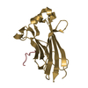
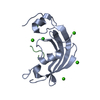
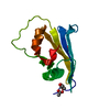
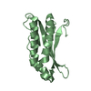
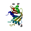
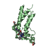
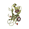
| Title | Ribosome Binding Factor A(rbfA) | ||||||
 Components Components | RIBOSOME-BINDING FACTOR A | ||||||
 Keywords Keywords | RNA BINDING PROTEIN / Structure 2 Function Project / S2F / Structural Genomics | ||||||
| Function / homology |  Function and homology information Function and homology informationribosomal small subunit binding / maturation of SSU-rRNA / ribosome biogenesis / cytosol Similarity search - Function | ||||||
| Biological species |  Haemophilus influenzae (bacteria) Haemophilus influenzae (bacteria) | ||||||
| Method |  X-RAY DIFFRACTION / SYNCHROTRON / MAD / Resolution: 1.7 Å X-RAY DIFFRACTION / SYNCHROTRON / MAD / Resolution: 1.7 Å | ||||||
 Authors Authors | Bonander, N. / Tordova, M. / Howard, A.J. / Eisenstein, E. / Gilliland, G.L. / Structure 2 Function Project (S2F) | ||||||
 Citation Citation | Journal: To be Published Title: The 1.7-A Crystal Structure of HI1288 - Ribosome Binding Factor A (rbfA), a Cold Response Protein Authors: Bonander, N. / Tordova, M. / Howard, A.J. / Eisenstein, E. / Gilliland, G.L. | ||||||
| History |
|
- Structure visualization
| Structure viewer | Molecule: MolmilJmol/JSmol |
|---|
- Downloads & links
-Download
| PDBx/mmCIF format | 1jos.cif.gz | 34.8 KB | Display | PDBx/mmCIF format |
|---|---|---|---|---|
| PDB format | pdb1jos.ent.gz | 23.7 KB | Display | PDB format |
| PDBx/mmJSON format | 1jos.json.gz | Tree view | PDBx/mmJSON format | |
| Others |  Other downloads Other downloads |
-Validation report
| Arichive directory | https://data.pdbj.org/pub/pdb/validation_reports/jo/1josftp://data.pdbj.org/pub/pdb/validation_reports/jo/1jos | HTTPS FTP |
|---|
-Related structure data
| Similar structure data | |
|---|---|
| Other databases |


-Links
 PDBj
PDBj- Assembly
| Deposited unit | 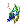
| ||||||||
|---|---|---|---|---|---|---|---|---|---|
| 1 |
| ||||||||
| Unit cell |
|
-Components
| #1: Protein | Mass: 14827.134 Da / Num. of mol.: 1 Source method: isolated from a genetically manipulated source Source: (gene. exp.) Haemophilus influenzae (bacteria) / Gene: HI1288 / Production host: |
|---|---|
| #2: Water | ChemComp-HOH /  Mass: 18.015 Da / Num. of mol.: 169 / Source method: isolated from a natural source / Formula: H2O Mass: 18.015 Da / Num. of mol.: 169 / Source method: isolated from a natural source / Formula: H2O |
-Experimental details
-Experiment
| Experiment | Method: X-RAY DIFFRACTION / Number of used crystals: 1 |
|---|
- Sample preparation
| Crystal | Density Matthews: 2.17 Å3/Da / Density % sol: 43.38 % |
|---|---|
| Crystal grow | Temperature: 293 K / Method: vapor diffusion, hanging drop / pH: 7.3 Details: 0.6M NaK tartrate, 10 mM HEPES, pH 7.3, VAPOR DIFFUSION, HANGING DROP, temperature 293K |
-Data collection
| Diffraction | Mean temperature: 115 K |
|---|---|
| Diffraction source | Source: SYNCHROTRON / Site: APS  / Beamline: 17-ID / Wavelength: 1.0113 Å / Beamline: 17-ID / Wavelength: 1.0113 Å |
| Detector | Type: BRUKER / Detector: CCD / Date: Jun 1, 2000 |
| Radiation | Protocol: SINGLE WAVELENGTH / Monochromatic (M) / Laue (L): M / Scattering type: x-ray |
| Radiation wavelength | Wavelength: 1.0113 Å / Relative weight: 1 |
| Reflection | Resolution: 1.7→15 Å / Num. all: 14727 / Num. obs: 10622 / % possible obs: 72 % / Rmerge(I) obs: 0.124 / Net I/σ(I): 12.7 |
| Reflection shell | Resolution: 1.7→1.74 Å / Mean I/σ(I) obs: 3.5 / Rsym value: 0.32 |
- Processing
| Software |
| |||||||||||||||
|---|---|---|---|---|---|---|---|---|---|---|---|---|---|---|---|---|
| Refinement | Method to determine structure: MAD / Resolution: 1.7→15 Å / Rfactor Rfree: 0.283 / Rfactor Rwork: 0.216 | |||||||||||||||
| Refinement step | Cycle: LAST / Resolution: 1.7→15 Å
| |||||||||||||||
| Refine LS restraints |
|
