 Movie
Movie Controller

[English] 日本語
 Yorodumi
- PDB-1jl4: CRYSTAL STRUCTURE OF THE HUMAN CD4 N-TERMINAL TWO DOMAIN FRAGMENT... -
+ Open data

- Basic information
| Entry | Database: PDB / ID: 1jl4 | ||||||
|---|---|---|---|---|---|---|---|
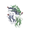
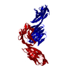
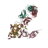
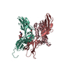
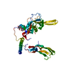
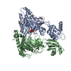
| Title | CRYSTAL STRUCTURE OF THE HUMAN CD4 N-TERMINAL TWO DOMAIN FRAGMENT COMPLEXED TO A CLASS II MHC MOLECULE | ||||||
 Components Components |
| ||||||
 Keywords Keywords |  IMMUNE SYSTEM / PROTEIN-PROTEIN COMPLEX IMMUNE SYSTEM / PROTEIN-PROTEIN COMPLEX | ||||||
| Function / homology |  Function and homology information Function and homology informationextracellular sequestering of iron ion / organomineral extracellular matrix / Phosphorylation of CD3 and TCR zeta chains / Translocation of ZAP-70 to Immunological synapse / PD-1 signaling / Generation of second messenger molecules / helper T cell enhancement of adaptive immune response / interleukin-16 binding / interleukin-16 receptor activity / Downstream TCR signaling ...extracellular sequestering of iron ion / organomineral extracellular matrix / Phosphorylation of CD3 and TCR zeta chains / Translocation of ZAP-70 to Immunological synapse / PD-1 signaling / Generation of second messenger molecules / helper T cell enhancement of adaptive immune response / interleukin-16 binding / interleukin-16 receptor activity / Downstream TCR signaling / maintenance of protein location in cell / T cell selection / antigen processing and presentation of peptide antigen / MHC class II protein binding / MHC class II antigen presentation / cellular response to granulocyte macrophage colony-stimulating factor stimulus / interleukin-15-mediated signaling pathway / positive regulation of monocyte differentiation / Nef Mediated CD4 Down-regulation / antimicrobial humoral response / Alpha-defensins / positive regulation of kinase activity / regulation of T cell activation / positive regulation of T cell differentiation / T cell receptor complex / extracellular matrix structural constituent / Other interleukin signaling / enzyme-linked receptor protein signaling pathway / Translocation of ZAP-70 to Immunological synapse / Phosphorylation of CD3 and TCR zeta chains / intracellular sequestering of iron ion / regulation of calcium ion transport / macrophage differentiation / Generation of second messenger molecules / T cell differentiation / antigen processing and presentation / PD-1 signaling / positive regulation of protein kinase activity / Binding and entry of HIV virion / coreceptor activity / positive regulation of calcium-mediated signaling / negative regulation of T cell proliferation / protein tyrosine kinase binding / positive regulation of interleukin-2 production / T cell activation / multivesicular body / ferric iron binding / Vpu mediated degradation of CD4 / calcium-mediated signaling / acute-phase response / clathrin-coated endocytic vesicle membrane / recycling endosome / peptide antigen assembly with MHC class II protein complex / MHC class II protein complex / peptide antigen binding / cell surface receptor protein tyrosine kinase signaling pathway / antigen processing and presentation of exogenous peptide antigen via MHC class II / positive regulation of immune response / positive regulation of T cell activation / positive regulation of peptidyl-tyrosine phosphorylation / transmembrane signaling receptor activity / Cargo recognition for clathrin-mediated endocytosis / Downstream TCR signaling / virus receptor activity / signaling receptor activity / Clathrin-mediated endocytosis / MHC class II protein complex binding / late endosome membrane / antibacterial humoral response / iron ion transport / positive regulation of canonical NF-kappaB signal transduction / defense response to Gram-negative bacterium / response to lipopolysaccharide / positive regulation of MAPK cascade / adaptive immune response / positive regulation of viral entry into host cell / lysosome / positive regulation of ERK1 and ERK2 cascade / cell surface receptor signaling pathway / early endosome / cell adhesion / immune response / response to xenobiotic stimulus / positive regulation of protein phosphorylation / iron ion binding / membrane raft / lysosomal membrane / endoplasmic reticulum lumen / external side of plasma membrane / lipid binding / protein-containing complex binding / endoplasmic reticulum membrane / protein kinase binding / positive regulation of DNA-templated transcription / Golgi apparatus / enzyme binding / cell surface / signal transduction / protein homodimerization activity / extracellular spaceSimilarity search - Function | ||||||
| Biological species |  Mus musculus (house mouse) Mus musculus (house mouse) Gallus gallus (chicken) Gallus gallus (chicken) Homo sapiens (human) Homo sapiens (human) | ||||||
| Method | X-RAY DIFFRACTION / SYNCHROTRON / MOLECULAR REPLACEMENT / Resolution: 4.3 Å | ||||||
 Authors Authors | Wang, J.-H. / Meijers, R. / Reinherz, E.L. | ||||||
 Citation Citation | Journal: Proc.Natl.Acad.Sci.USA / Year: 2001 Title: Crystal structure of the human CD4 N-terminal two-domain fragment complexed to a class II MHC molecule. Authors: Wang, J.H. / Meijers, R. / Xiong, Y. / Liu, J.H. / Sakihama, T. / Zhang, R. / Joachimiak, A. / Reinherz, E.L. | ||||||
| History |
|
- Structure visualization
| Structure viewer | Molecule: MolmilJmol/JSmol |
|---|
- Downloads & links
-Download
| PDBx/mmCIF format | 1jl4.cif.gz | 112.3 KB | Display | PDBx/mmCIF format |
|---|---|---|---|---|
| PDB format | pdb1jl4.ent.gz | 83.9 KB | Display | PDB format |
| PDBx/mmJSON format | 1jl4.json.gz | Tree view | PDBx/mmJSON format | |
| Others |  Other downloads Other downloads |
-Validation report
| Arichive directory | https://data.pdbj.org/pub/pdb/validation_reports/jl/1jl4ftp://data.pdbj.org/pub/pdb/validation_reports/jl/1jl4 | HTTPS FTP |
|---|
-Related structure data


-Links
 PDBj
PDBj


- Assembly
| Deposited unit | 
| ||||||||
|---|---|---|---|---|---|---|---|---|---|
| 1 |
| ||||||||
| Unit cell |
|
-Components
| #1: Protein | Mass: 20429.812 Da / Num. of mol.: 1 Source method: isolated from a genetically manipulated source Source: (gene. exp.) Mus musculus (house mouse) / Production host:  Cricetulus griseus (Chinese hamster) / References: UniProt: P01910 Cricetulus griseus (Chinese hamster) / References: UniProt: P01910 |
|---|---|
| #2: Protein | Mass: 21887.592 Da / Num. of mol.: 1 Source method: isolated from a genetically manipulated source Source: (gene. exp.) Mus musculus (house mouse) / Production host: Cricetulus griseus (Chinese hamster) / References: UniProt: P06343 |
| #3: Protein/peptide | / CONALBUMIN Mass: 1700.766 Da / Num. of mol.: 1 Source method: isolated from a genetically manipulated source Source: (gene. exp.) Gallus gallus (chicken) / Production host: Gallus gallus (chicken) / References: UniProt: P02789 |
| #4: Protein | Mass: 19725.414 Da / Num. of mol.: 1 Source method: isolated from a genetically manipulated source Source: (gene. exp.) Homo sapiens (human) / Production host: Cricetulus griseus (Chinese hamster) / References: UniProt: P01730 |
-Experimental details
-Experiment
| Experiment | Method: X-RAY DIFFRACTION / Number of used crystals: 1 |
|---|
- Sample preparation
| Crystal | Density Matthews: 4.29 Å3/Da / Density % sol: 70 % | ||||||||||||||||||||||||||||||
|---|---|---|---|---|---|---|---|---|---|---|---|---|---|---|---|---|---|---|---|---|---|---|---|---|---|---|---|---|---|---|---|
| Crystal grow | Temperature: 297 K / Method: vapor diffusion, hanging drop / pH: 8.5 Details: 17% PEG 4,000/0.2M Li2SO4/0.1M Tris, pH 8.5, VAPOR DIFFUSION, HANGING DROP, temperature 297K | ||||||||||||||||||||||||||||||
| Crystal grow | *PLUS | ||||||||||||||||||||||||||||||
| Components of the solutions | *PLUS
|
-Data collection
| Diffraction | Mean temperature: 100 K |
|---|---|
| Diffraction source | Source: SYNCHROTRON / Site: APS  / Beamline: 19-ID / Wavelength: 1.033 Å / Beamline: 19-ID / Wavelength: 1.033 Å |
| Detector | Type: SBC-2 / Detector: CCD / Date: Dec 12, 2000 / Details: mirrors |
| Radiation | Monochromator: Si 111 / Protocol: SINGLE WAVELENGTH / Monochromatic (M) / Laue (L): M / Scattering type: x-ray |
| Radiation wavelength | Wavelength: 1.033 Å / Relative weight: 1 |
| Reflection | Resolution: 4.3→30 Å / Num. all: 7428 / Num. obs: 7428 / % possible obs: 83 % / Observed criterion σ(F): 0 / Observed criterion σ(I): 0 / Rmerge(I) obs: 0.152 / Net I/σ(I): 12 |
| Reflection shell | Resolution: 4.3→4.5 Å / Redundancy: 7.8 % / Rmerge(I) obs: 0.61 / Mean I/σ(I) obs: 2 / % possible all: 56 |
| Reflection | *PLUS % possible obs: 83 % / Redundancy: 9.6 % |
| Reflection shell | *PLUS % possible obs: 56 % |
- Processing
| Software |
| |||||||||||||||||||||||||
|---|---|---|---|---|---|---|---|---|---|---|---|---|---|---|---|---|---|---|---|---|---|---|---|---|---|---|
| Refinement | Method to determine structure: MOLECULAR REPLACEMENT Starting model: 1IAK and 3CD4 Resolution: 4.3→20 Å / σ(F): 0 / Stereochemistry target values: Engh & Huber Details: THIS MODEL IS BASED ON HIGH RESOLUTION STRUCTURES OF THE INDIVIDUAL COMPONENTS AND ONLY RIGID BODY REFINEMENT OF THE INDIVIDUAL DOMAINS WAS APPLIED. THE RIGID BODIES CONSISTED OF RESIDUE ...Details: THIS MODEL IS BASED ON HIGH RESOLUTION STRUCTURES OF THE INDIVIDUAL COMPONENTS AND ONLY RIGID BODY REFINEMENT OF THE INDIVIDUAL DOMAINS WAS APPLIED. THE RIGID BODIES CONSISTED OF RESIDUE RANGES A 5 TO A 85, A 86 TO A 181, B 5 TO B 85, B 86 TO B 190, C 131 TO C 146, D 1 TO D 97 AND D 98 TO D 178
| |||||||||||||||||||||||||
| Refinement step | Cycle: LAST / Resolution: 4.3→20 Å
| |||||||||||||||||||||||||
| LS refinement shell | Resolution: 4.3→4.5 Å
| |||||||||||||||||||||||||
| Software | *PLUS Name: REFMAC / Classification: refinement | |||||||||||||||||||||||||
| Refinement | *PLUS Highest resolution: 4.3 Å / σ(F): 0 / Rfactor Rwork: 0.42 | |||||||||||||||||||||||||
| Solvent computation | *PLUS | |||||||||||||||||||||||||
| Displacement parameters | *PLUS | |||||||||||||||||||||||||
| Refine LS restraints | *PLUS
|
