 Movie
Movie Controller


+ Open data

- Basic information
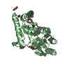
| Entry | Database: PDB / ID: 1jjv | ||||||
|---|---|---|---|---|---|---|---|
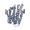
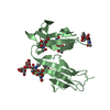
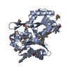
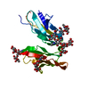
| Title | DEPHOSPHO-COA KINASE IN COMPLEX WITH ATP | ||||||
 Components Components | DEPHOSPHO-COA KINASE | ||||||
 Keywords Keywords | TRANSFERASE / P-loop nucleotide-binding fold / Structure 2 Function Project / S2F / Structural Genomics | ||||||
| Function / homology |  Function and homology informationdephospho-CoA kinase / dephospho-CoA kinase activity / coenzyme A biosynthetic process / ATP binding / cytoplasm Function and homology informationdephospho-CoA kinase / dephospho-CoA kinase activity / coenzyme A biosynthetic process / ATP binding / cytoplasmSimilarity search - Function | ||||||
| Biological species |  Haemophilus influenzae (bacteria) Haemophilus influenzae (bacteria) | ||||||
| Method | X-RAY DIFFRACTION / SYNCHROTRON / MAD / Resolution: 2 Å | ||||||
 Authors Authors | Obmolova, G. / Teplyakov, A. / Bonander, N. / Eisenstein, E. / Howard, A.J. / Gilliland, G.L. / Structure 2 Function Project (S2F) | ||||||
 Citation Citation | Journal: J.Struct.Biol. / Year: 2001 Title: Crystal structure of dephospho-coenzyme A kinase from Haemophilus influenzae. Authors: Obmolova, G. / Teplyakov, A. / Bonander, N. / Eisenstein, E. / Howard, A.J. / Gilliland, G.L. | ||||||
| History |
|
- Structure visualization
| Structure viewer | Molecule: MolmilJmol/JSmol |
|---|
- Downloads & links
-Download
| PDBx/mmCIF format | 1jjv.cif.gz | 57.9 KB | Display | PDBx/mmCIF format |
|---|---|---|---|---|
| PDB format | pdb1jjv.ent.gz | 41.1 KB | Display | PDB format |
| PDBx/mmJSON format | 1jjv.json.gz | Tree view | PDBx/mmJSON format | |
| Others |  Other downloads Other downloads |
-Validation report
| Arichive directory | https://data.pdbj.org/pub/pdb/validation_reports/jj/1jjvftp://data.pdbj.org/pub/pdb/validation_reports/jj/1jjv | HTTPS FTP |
|---|
-Related structure data
| Similar structure data | |
|---|---|
| Other databases |


-Links
 PDBj
PDBj


- Assembly
| Deposited unit | 
| ||||||||
|---|---|---|---|---|---|---|---|---|---|
| 1 |
| ||||||||
| Unit cell |
|
-Components
| #1: Protein | / DEPHOSPHOCOENZYME A KINASE Mass: 23312.502 Da / Num. of mol.: 1 Source method: isolated from a genetically manipulated source Source: (gene. exp.) Haemophilus influenzae (bacteria) / Gene: YacE / Plasmid: pET15b / Species (production host): Escherichia coli / Production host: Escherichia coli BL21 (bacteria) / Strain (production host): BL21 / References: UniProt: P44920, dephospho-CoA kinase |
|---|---|
| #2: Chemical | ChemComp-SO4 / Sulfate  Mass: 96.063 Da / Num. of mol.: 1 / Source method: obtained synthetically / Formula: SO4 Mass: 96.063 Da / Num. of mol.: 1 / Source method: obtained synthetically / Formula: SO4 |
| #3: Chemical | ChemComp-HG / Mercury (element)  Mass: 200.590 Da / Num. of mol.: 1 / Source method: obtained synthetically / Formula: Hg Mass: 200.590 Da / Num. of mol.: 1 / Source method: obtained synthetically / Formula: Hg |
| #4: Chemical | ChemComp-ATP / Adenosine triphosphate  Mass: 507.181 Da / Num. of mol.: 1 / Source method: obtained synthetically / Formula: C10H16N5O13P3 / Comment: ATP, energy-carrying molecule*YM Mass: 507.181 Da / Num. of mol.: 1 / Source method: obtained synthetically / Formula: C10H16N5O13P3 / Comment: ATP, energy-carrying molecule*YM |
| #5: Water | ChemComp-HOH / Water Mass: 18.015 Da / Num. of mol.: 207 / Source method: isolated from a natural source / Formula: H2O Mass: 18.015 Da / Num. of mol.: 207 / Source method: isolated from a natural source / Formula: H2O |
-Experimental details
-Experiment
| Experiment | Method: X-RAY DIFFRACTION / Number of used crystals: 1 |
|---|
- Sample preparation
| Crystal | Density Matthews: 2.58 Å3/Da / Density % sol: 52.1 % | |||||||||||||||||||||||||||||||||||||||||||||||||||||||||||||||
|---|---|---|---|---|---|---|---|---|---|---|---|---|---|---|---|---|---|---|---|---|---|---|---|---|---|---|---|---|---|---|---|---|---|---|---|---|---|---|---|---|---|---|---|---|---|---|---|---|---|---|---|---|---|---|---|---|---|---|---|---|---|---|---|---|
| Crystal grow | Temperature: 300 K / Method: vapor diffusion, hanging drop / pH: 7 Details: ammonium sulfate, PEG 400, pH 7, VAPOR DIFFUSION, HANGING DROP, temperature 300K | |||||||||||||||||||||||||||||||||||||||||||||||||||||||||||||||
| Crystal grow | *PLUS pH: 7 / Details: used microseeding | |||||||||||||||||||||||||||||||||||||||||||||||||||||||||||||||
| Components of the solutions | *PLUS
|
-Data collection
| Diffraction | Mean temperature: 100 K |
|---|---|
| Diffraction source | Source: SYNCHROTRON / Site: APS  / Beamline: 17-ID / Wavelength: 0.9999 Å / Beamline: 17-ID / Wavelength: 0.9999 Å |
| Detector | Type: MARRESEARCH / Detector: CCD / Date: Jun 17, 2001 |
| Radiation | Monochromator: SI(111) / Protocol: MAD / Monochromatic (M) / Laue (L): M / Scattering type: x-ray |
| Radiation wavelength | Wavelength: 0.9999 Å / Relative weight: 1 |
| Reflection | Resolution: 2→50 Å / Num. all: 15767 / Num. obs: 15767 / % possible obs: 98 % / Observed criterion σ(F): 0 / Observed criterion σ(I): 0 / Redundancy: 3.6 % / Biso Wilson estimate: 25.6 Å2 / Rsym value: 0.057 / Net I/σ(I): 37.2 |
| Reflection shell | Resolution: 2→2.05 Å / Redundancy: 2.6 % / Mean I/σ(I) obs: 7.5 / Rsym value: 0.121 / % possible all: 83 |
| Reflection | *PLUS Highest resolution: 2 Å / Lowest resolution: 50 Å / % possible obs: 97.6 % / Num. measured all: 56478 / Rmerge(I) obs: 0.057 |
- Processing
| Software |
| |||||||||||||||||||||||||||||||||||||||||||||||||||||||||||||||
|---|---|---|---|---|---|---|---|---|---|---|---|---|---|---|---|---|---|---|---|---|---|---|---|---|---|---|---|---|---|---|---|---|---|---|---|---|---|---|---|---|---|---|---|---|---|---|---|---|---|---|---|---|---|---|---|---|---|---|---|---|---|---|---|---|
| Refinement | Method to determine structure: MAD / Resolution: 2→10 Å / SU B: 3.6 / SU ML: 0.1 / σ(F): 0 / ESU R: 0.16 / ESU R Free: 0.15 / Stereochemistry target values: Engh & Huber
| |||||||||||||||||||||||||||||||||||||||||||||||||||||||||||||||
| Refine analyze |
| |||||||||||||||||||||||||||||||||||||||||||||||||||||||||||||||
| Refinement step | Cycle: LAST / Resolution: 2→10 Å
| |||||||||||||||||||||||||||||||||||||||||||||||||||||||||||||||
| Refine LS restraints |
| |||||||||||||||||||||||||||||||||||||||||||||||||||||||||||||||
| Refinement | *PLUS Highest resolution: 2 Å / Lowest resolution: 10 Å / % reflection Rfree: 5 % / Rfactor Rwork: 0.168 | |||||||||||||||||||||||||||||||||||||||||||||||||||||||||||||||
| Solvent computation | *PLUS | |||||||||||||||||||||||||||||||||||||||||||||||||||||||||||||||
| Displacement parameters | *PLUS | |||||||||||||||||||||||||||||||||||||||||||||||||||||||||||||||
| Refine LS restraints | *PLUS
|
