 Movie
Movie Controller

[English] 日本語
 Yorodumi
- PDB-7akx: Crystal structure of the viral rhodopsin OLPVR1 in P1 space group -
+ Open data

- Basic information
| Entry | Database: PDB / ID: 7akx | ||||||||||||
|---|---|---|---|---|---|---|---|---|---|---|---|---|---|
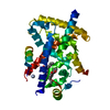
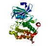
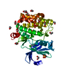
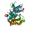
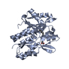
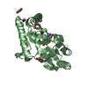
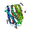
| Title | Crystal structure of the viral rhodopsin OLPVR1 in P1 space group | ||||||||||||
 Components Components | viral rhodopsin OLPVR1 | ||||||||||||
 Keywords Keywords | MEMBRANE PROTEIN / rhodopsin / viral rhodopsin / giant virus / ion channel / retinal / ion transport / light-gated channel / channelrhodopsin / bacteriorhodopsin | ||||||||||||
| Function / homology |  Function and homology information Function and homology informationphotoreceptor activity / phototransduction / monoatomic ion channel activity / membrane Similarity search - Function | ||||||||||||
| Biological species |  Organic Lake phycodnavirus (environmental samples) Organic Lake phycodnavirus (environmental samples) | ||||||||||||
| Method |  X-RAY DIFFRACTION / SYNCHROTRON / MOLECULAR REPLACEMENT / Resolution: 1.6 Å X-RAY DIFFRACTION / SYNCHROTRON / MOLECULAR REPLACEMENT / Resolution: 1.6 Å | ||||||||||||
 Authors Authors | Kovalev, K. / Zabelskii, D. / Alekseev, A. / Astashkin, R. / Gordeliy, V. | ||||||||||||
| Funding support |  France, France,  Russian Federation, 3items Russian Federation, 3items
| ||||||||||||
 Citation Citation | Journal: Nat Commun / Year: 2020 Title: Viral rhodopsins 1 are an unique family of light-gated cation channels. Authors: Zabelskii, D. / Alekseev, A. / Kovalev, K. / Rankovic, V. / Balandin, T. / Soloviov, D. / Bratanov, D. / Savelyeva, E. / Podolyak, E. / Volkov, D. / Vaganova, S. / Astashkin, R. / Chizhov, I. ...Authors: Zabelskii, D. / Alekseev, A. / Kovalev, K. / Rankovic, V. / Balandin, T. / Soloviov, D. / Bratanov, D. / Savelyeva, E. / Podolyak, E. / Volkov, D. / Vaganova, S. / Astashkin, R. / Chizhov, I. / Yutin, N. / Rulev, M. / Popov, A. / Eria-Oliveira, A.S. / Rokitskaya, T. / Mager, T. / Antonenko, Y. / Rosselli, R. / Armeev, G. / Shaitan, K. / Vivaudou, M. / Buldt, G. / Rogachev, A. / Rodriguez-Valera, F. / Kirpichnikov, M. / Moser, T. / Offenhausser, A. / Willbold, D. / Koonin, E. / Bamberg, E. / Gordeliy, V. | ||||||||||||
| History |
|
- Structure visualization
| Structure viewer | Molecule: MolmilJmol/JSmol |
|---|
- Downloads & links
-Download
| PDBx/mmCIF format | 7akx.cif.gz | 123.3 KB | Display | PDBx/mmCIF format |
|---|---|---|---|---|
| PDB format | pdb7akx.ent.gz | 92.7 KB | Display | PDB format |
| PDBx/mmJSON format | 7akx.json.gz | Tree view | PDBx/mmJSON format | |
| Others |  Other downloads Other downloads |
-Validation report
| Arichive directory | https://data.pdbj.org/pub/pdb/validation_reports/ak/7akxftp://data.pdbj.org/pub/pdb/validation_reports/ak/7akx | HTTPS FTP |
|---|
-Related structure data
| Related structure data |  7akwC  7akyC  6sqgS S: Starting model for refinement C: citing same article ( |
|---|---|
| Similar structure data |


-Links
 PDBj
PDBj


- Assembly
| Deposited unit | 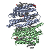
| ||||||||
|---|---|---|---|---|---|---|---|---|---|
| 1 | 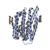
| ||||||||
| 2 | 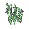
| ||||||||
| Unit cell |
|
-Components
| #1: Protein | Mass: 28366.715 Da / Num. of mol.: 2 Source method: isolated from a genetically manipulated source Source: (gene. exp.) Organic Lake phycodnavirus (environmental samples)Gene: 162281038 / Production host:  #2: Chemical | ChemComp-LFA /   Mass: 282.547 Da / Num. of mol.: 26 / Source method: obtained synthetically / Formula: C20H42 Mass: 282.547 Da / Num. of mol.: 26 / Source method: obtained synthetically / Formula: C20H42#3: Chemical | ChemComp-OLA /   Mass: 282.461 Da / Num. of mol.: 4 / Source method: obtained synthetically / Formula: C18H34O2 Mass: 282.461 Da / Num. of mol.: 4 / Source method: obtained synthetically / Formula: C18H34O2#4: Chemical |   Mass: 356.540 Da / Num. of mol.: 2 / Source method: obtained synthetically / Formula: C21H40O4 Mass: 356.540 Da / Num. of mol.: 2 / Source method: obtained synthetically / Formula: C21H40O4#5: Water | ChemComp-HOH / |  Mass: 18.015 Da / Num. of mol.: 98 / Source method: isolated from a natural source / Formula: H2O Mass: 18.015 Da / Num. of mol.: 98 / Source method: isolated from a natural source / Formula: H2OHas ligand of interest | Y | |
|---|
-Experimental details
-Experiment
| Experiment | Method: X-RAY DIFFRACTION / Number of used crystals: 1 |
|---|
- Sample preparation
| Crystal | Density Matthews: 2.39 Å3/Da / Density % sol: 48.57 % |
|---|---|
| Crystal grow | Temperature: 293 K / Method: lipidic cubic phase / pH: 8.2 Details: 10 mM CaCl2, 10 mM MgCl2, 24% PEG 550, 100mM Tris (pH 8.2), monoolein |
-Data collection
| Diffraction | Mean temperature: 100 K / Serial crystal experiment: N | ||||||||||||||||||||||||||||||
|---|---|---|---|---|---|---|---|---|---|---|---|---|---|---|---|---|---|---|---|---|---|---|---|---|---|---|---|---|---|---|---|
| Diffraction source | Source: SYNCHROTRON / Site: ESRF / Beamline: ID30B / Wavelength: 0.97625 Å | ||||||||||||||||||||||||||||||
| Detector | Type: DECTRIS PILATUS 6M-F / Detector: PIXEL / Date: Jun 6, 2018 | ||||||||||||||||||||||||||||||
| Radiation | Protocol: SINGLE WAVELENGTH / Monochromatic (M) / Laue (L): M / Scattering type: x-ray | ||||||||||||||||||||||||||||||
| Radiation wavelength | Wavelength: 0.97625 Å / Relative weight: 1 | ||||||||||||||||||||||||||||||
| Reflection | Resolution: 1.6→40.16 Å / Num. obs: 63486 / % possible obs: 95.2 % / Redundancy: 3.5 % / CC1/2: 0.998 / Rmerge(I) obs: 0.058 / Rpim(I) all: 0.036 / Rrim(I) all: 0.069 / Net I/σ(I): 6.1 / Num. measured all: 221715 | ||||||||||||||||||||||||||||||
| Reflection shell | Diffraction-ID: 1
|
- Processing
| Software |
| ||||||||||||||||||||||||||||||||||||||||||||||||||||||||||||
|---|---|---|---|---|---|---|---|---|---|---|---|---|---|---|---|---|---|---|---|---|---|---|---|---|---|---|---|---|---|---|---|---|---|---|---|---|---|---|---|---|---|---|---|---|---|---|---|---|---|---|---|---|---|---|---|---|---|---|---|---|---|
| Refinement | Method to determine structure: MOLECULAR REPLACEMENT Starting model: 6sqg Resolution: 1.6→20 Å / Cor.coef. Fo:Fc: 0.965 / Cor.coef. Fo:Fc free: 0.959 / SU B: 2.711 / SU ML: 0.086 / Cross valid method: THROUGHOUT / σ(F): 0 / ESU R: 0.099 / ESU R Free: 0.095 / Stereochemistry target values: MAXIMUM LIKELIHOOD Details: HYDROGENS HAVE BEEN ADDED IN THE RIDING POSITIONS U VALUES : REFINED INDIVIDUALLY
| ||||||||||||||||||||||||||||||||||||||||||||||||||||||||||||
| Solvent computation | Ion probe radii: 0.8 Å / Shrinkage radii: 0.8 Å / VDW probe radii: 1.2 Å / Solvent model: MASK | ||||||||||||||||||||||||||||||||||||||||||||||||||||||||||||
| Displacement parameters | Biso max: 117.37 Å2 / Biso mean: 29.79 Å2 / Biso min: 16.67 Å2
| ||||||||||||||||||||||||||||||||||||||||||||||||||||||||||||
| Refinement step | Cycle: final / Resolution: 1.6→20 Å
| ||||||||||||||||||||||||||||||||||||||||||||||||||||||||||||
| Refine LS restraints |
| ||||||||||||||||||||||||||||||||||||||||||||||||||||||||||||
| LS refinement shell | Resolution: 1.6→1.641 Å / Rfactor Rfree error: 0 / Total num. of bins used: 20
|
