 Movie
Movie Controller

[English] 日本語
 Yorodumi
- PDB-1jgi: Crystal Structure of the Active Site Mutant Glu328Gln of Amylosuc... -
+ Open data

- Basic information
| Entry | Database: PDB / ID: 1jgi | |||||||||
|---|---|---|---|---|---|---|---|---|---|---|
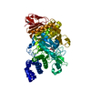
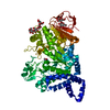
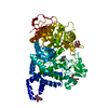
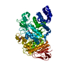
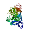
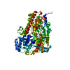
| Title | Crystal Structure of the Active Site Mutant Glu328Gln of Amylosucrase from Neisseria polysaccharea in Complex with the Natural Substrate Sucrose | |||||||||
 Components Components | amylosucrase | |||||||||
 Keywords Keywords | TRANSFERASE / active site mutant Glu328Gln / sucrose complex | |||||||||
| Function / homology |  Function and homology information Function and homology informationamylosucrase / amylosucrase activity / carbohydrate metabolic process / extracellular region Similarity search - Function | |||||||||
| Biological species |  Neisseria polysaccharea (bacteria) Neisseria polysaccharea (bacteria) | |||||||||
| Method |  X-RAY DIFFRACTION / SYNCHROTRON / MOLECULAR REPLACEMENT / Resolution: 2 Å X-RAY DIFFRACTION / SYNCHROTRON / MOLECULAR REPLACEMENT / Resolution: 2 Å | |||||||||
 Authors Authors | Mirza, O. / Skov, L.K. / Gajhede, M. | |||||||||
 Citation Citation | Journal: Biochemistry / Year: 2001 Title: Crystal structures of amylosucrase from Neisseria polysaccharea in complex with D-glucose and the active site mutant Glu328Gln in complex with the natural substrate sucrose. Authors: Mirza, O. / Skov, L.K. / Remaud-Simeon, M. / Potocki de Montalk, G. / Albenne, C. / Monsan, P. / Gajhede, M. | |||||||||
| History |
| |||||||||
| Remark 999 | SEQUENCE THE DISCREPANCY BETWEEN RESIDUES TYR 131 & ASP 537 AND THE GENBANK SEQUENCE DATABASE ...SEQUENCE THE DISCREPANCY BETWEEN RESIDUES TYR 131 & ASP 537 AND THE GENBANK SEQUENCE DATABASE REFERENCE, ACCESSION 4107260, RESIDUES HIS 139 & GLY 545, RESPECTIVELY, IS DUE TO A PCR ERROR. |
- Structure visualization
| Structure viewer | Molecule: MolmilJmol/JSmol |
|---|
- Downloads & links
-Download
| PDBx/mmCIF format | 1jgi.cif.gz | 149.9 KB | Display | PDBx/mmCIF format |
|---|---|---|---|---|
| PDB format | pdb1jgi.ent.gz | 115.8 KB | Display | PDB format |
| PDBx/mmJSON format | 1jgi.json.gz | Tree view | PDBx/mmJSON format | |
| Others |  Other downloads Other downloads |
-Validation report
| Summary document | 1jgi_validation.pdf.gz | 438.5 KB | Display | wwPDB validaton report |
|---|---|---|---|---|
| Full document | 1jgi_full_validation.pdf.gz | 447.6 KB | Display | |
| Data in XML | 1jgi_validation.xml.gz | 14.1 KB | Display | |
| Data in CIF | 1jgi_validation.cif.gz | 24.5 KB | Display | |
| Arichive directory | https://data.pdbj.org/pub/pdb/validation_reports/jg/1jgiftp://data.pdbj.org/pub/pdb/validation_reports/jg/1jgi | HTTPS FTP |
-Related structure data


-Links
 PDBj
PDBj

- Assembly
| Deposited unit | 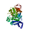
| ||||||||
|---|---|---|---|---|---|---|---|---|---|
| 1 |
| ||||||||
| Unit cell |
|
-Components
| #1: Protein | Mass: 71588.117 Da / Num. of mol.: 1 / Mutation: E328Q Source method: isolated from a genetically manipulated source Source: (gene. exp.) Neisseria polysaccharea (bacteria) / Production host: |
|---|---|
| #2: Polysaccharide | beta-D-fructofuranose-(2-1)-alpha-D-glucopyranose / sucrose  Source method: isolated from a genetically manipulated source Details: oligosaccharide with reducing-end-to-reducing-end glycosidic bond References: sucrose |
| #3: Water | ChemComp-HOH /  Mass: 18.015 Da / Num. of mol.: 622 / Source method: isolated from a natural source / Formula: H2O Mass: 18.015 Da / Num. of mol.: 622 / Source method: isolated from a natural source / Formula: H2O |
-Experimental details
-Experiment
| Experiment | Method: X-RAY DIFFRACTION / Number of used crystals: 1 |
|---|
- Sample preparation
| Crystal | Density Matthews: 2.37 Å3/Da / Density % sol: 48.09 % | ||||||||||||||||||||||||||||||||||||||||||||||||
|---|---|---|---|---|---|---|---|---|---|---|---|---|---|---|---|---|---|---|---|---|---|---|---|---|---|---|---|---|---|---|---|---|---|---|---|---|---|---|---|---|---|---|---|---|---|---|---|---|---|
| Crystal grow | *PLUS Temperature: 4 ℃ / pH: 7 / Method: vapor diffusion, hanging drop | ||||||||||||||||||||||||||||||||||||||||||||||||
| Components of the solutions | *PLUS
|
-Data collection
| Diffraction | Mean temperature: 120 K |
|---|---|
| Diffraction source | Source: SYNCHROTRON / Site: MAX II  / Beamline: I711 / Wavelength: 1.106 Å / Beamline: I711 / Wavelength: 1.106 Å |
| Detector | Type: MARRESEARCH / Detector: CCD |
| Radiation | Protocol: SINGLE WAVELENGTH / Monochromatic (M) / Laue (L): M / Scattering type: x-ray |
| Radiation wavelength | Wavelength: 1.106 Å / Relative weight: 1 |
| Reflection | Resolution: 2→3 Å / Num. all: 335382 / Num. obs: 39254 / % possible obs: 84.2 % / Redundancy: 8 % / Biso Wilson estimate: 5.5 Å2 / Rmerge(I) obs: 0.076 / Net I/σ(I): 10 |
| Reflection shell | Resolution: 2→2.07 Å / Redundancy: 8 % / Rmerge(I) obs: 0.197 / Num. unique all: 3974 / % possible all: 86.8 |
| Reflection shell | *PLUS % possible obs: 86.8 % |
- Processing
| Software |
| ||||||||||||||||||||||||||||||||||||||||
|---|---|---|---|---|---|---|---|---|---|---|---|---|---|---|---|---|---|---|---|---|---|---|---|---|---|---|---|---|---|---|---|---|---|---|---|---|---|---|---|---|---|
| Refinement | Method to determine structure: MOLECULAR REPLACEMENT / Resolution: 2→27.91 Å / Rfactor Rfree error: 0.005 / Data cutoff high absF: 953468.23 / Data cutoff low absF: 0 / Isotropic thermal model: RESTRAINED / Cross valid method: THROUGHOUT / σ(F): 0
| ||||||||||||||||||||||||||||||||||||||||
| Solvent computation | Solvent model: FLAT MODEL / Bsol: 42.031 Å2 / ksol: 0.338146 e/Å3 | ||||||||||||||||||||||||||||||||||||||||
| Displacement parameters | Biso mean: 12.6 Å2
| ||||||||||||||||||||||||||||||||||||||||
| Refine analyze |
| ||||||||||||||||||||||||||||||||||||||||
| Refinement step | Cycle: LAST / Resolution: 2→27.91 Å
| ||||||||||||||||||||||||||||||||||||||||
| Refine LS restraints |
| ||||||||||||||||||||||||||||||||||||||||
| LS refinement shell | Resolution: 2→2.13 Å / Rfactor Rfree error: 0.014 / Total num. of bins used: 6
| ||||||||||||||||||||||||||||||||||||||||
| Xplor file |
| ||||||||||||||||||||||||||||||||||||||||
| Software | *PLUS Name: CNS / Version: 1 / Classification: refinement | ||||||||||||||||||||||||||||||||||||||||
| Refinement | *PLUS σ(F): 0 / % reflection Rfree: 5 % / Rfactor obs: 0.186 | ||||||||||||||||||||||||||||||||||||||||
| Solvent computation | *PLUS | ||||||||||||||||||||||||||||||||||||||||
| Displacement parameters | *PLUS Biso mean: 12.6 Å2 | ||||||||||||||||||||||||||||||||||||||||
| Refine LS restraints | *PLUS
| ||||||||||||||||||||||||||||||||||||||||
| LS refinement shell | *PLUS Rfactor Rfree: 0.26 / % reflection Rfree: 5.2 % / Rfactor Rwork: 0.214 |
