 Movie
Movie Controller

[English] 日本語
 Yorodumi
- PDB-4fls: Crystal structure of Amylosucrase inactive double mutant F290K-E3... -
+ Open data

- Basic information
| Entry | Database: PDB / ID: 4fls | |||||||||
|---|---|---|---|---|---|---|---|---|---|---|
| Title | Crystal structure of Amylosucrase inactive double mutant F290K-E328Q from Neisseria polysaccharea in complex with sucrose. | |||||||||
 Components Components | Amylosucrase | |||||||||
 Keywords Keywords | TRANSFERASE / BETA/ALPHA-BARREL / GLYCOSIDE HYDROLASE / AMYLOSE SYNTHESIS / SUCROSE ISOMERIZATION / GLUCOSYLTRANSFERASE CARBOHYDRATE | |||||||||
| Function / homology |  Function and homology information Function and homology informationamylosucrase / amylosucrase activity / carbohydrate metabolic process / extracellular region Similarity search - Function | |||||||||
| Biological species |  Neisseria polysaccharea (bacteria) Neisseria polysaccharea (bacteria) | |||||||||
| Method |  X-RAY DIFFRACTION / SYNCHROTRON / MOLECULAR REPLACEMENT / Resolution: 2.3 Å X-RAY DIFFRACTION / SYNCHROTRON / MOLECULAR REPLACEMENT / Resolution: 2.3 Å | |||||||||
 Authors Authors | Guerin, F. / Champion, E. / Moulis, C. / Barbe, S. / Tran, T.H. / Morel, S. / Descroix, K. / Monsan, P. / Mulard, L.A. / Remaud-Simeon, M. ...Guerin, F. / Champion, E. / Moulis, C. / Barbe, S. / Tran, T.H. / Morel, S. / Descroix, K. / Monsan, P. / Mulard, L.A. / Remaud-Simeon, M. / Andre, I. / Mourey, L. / Tranier, S. | |||||||||
 Citation Citation | Journal: J.Am.Chem.Soc. / Year: 2012 Title: Applying pairwise combinations of amino Acid mutations for sorting out highly efficient glucosylation tools for chemo-enzymatic synthesis of bacterial oligosaccharides. Authors: Champion, E. / Guerin, F. / Moulis, C. / Barbe, S. / Tran, T.H. / Morel, S. / Descroix, K. / Monsan, P. / Mourey, L. / Mulard, L.A. / Tranier, S. / Remaud-Simeon, M. / Andre, I. | |||||||||
| History |
|
- Structure visualization
| Structure viewer | Molecule: MolmilJmol/JSmol |
|---|
- Downloads & links
-Download
| PDBx/mmCIF format | 4fls.cif.gz | 143.8 KB | Display | PDBx/mmCIF format |
|---|---|---|---|---|
| PDB format | pdb4fls.ent.gz | 110.2 KB | Display | PDB format |
| PDBx/mmJSON format | 4fls.json.gz | Tree view | PDBx/mmJSON format | |
| Others |  Other downloads Other downloads |
-Validation report
| Arichive directory | https://data.pdbj.org/pub/pdb/validation_reports/fl/4flsftp://data.pdbj.org/pub/pdb/validation_reports/fl/4fls | HTTPS FTP |
|---|
-Related structure data
| Related structure data |  4floC  4flqC  4flrC  1g5aS S: Starting model for refinement C: citing same article ( |
|---|---|
| Similar structure data |


-Links
 PDBj
PDBj

- Assembly
| Deposited unit | 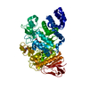
| ||||||||
|---|---|---|---|---|---|---|---|---|---|
| 1 |
| ||||||||
| Unit cell |
|
-Components
| #1: Protein | Mass: 71545.102 Da / Num. of mol.: 1 / Fragment: Amylosucrase (unp residues 13-636) / Mutation: F290K, E328Q Source method: isolated from a genetically manipulated source Source: (gene. exp.) Neisseria polysaccharea (bacteria) / Gene: ams / Plasmid: pGEX6p3 / Production host: | ||||||
|---|---|---|---|---|---|---|---|
| #2: Polysaccharide |   Source method: isolated from a genetically manipulated source Details: oligosaccharide with reducing-end-to-reducing-end glycosidic bond References: sucrose #3: Chemical | ChemComp-CL / |   Mass: 35.453 Da / Num. of mol.: 1 / Source method: obtained synthetically / Formula: Cl Mass: 35.453 Da / Num. of mol.: 1 / Source method: obtained synthetically / Formula: Cl#4: Chemical | ChemComp-GOL / |   Mass: 92.094 Da / Num. of mol.: 1 / Source method: obtained synthetically / Formula: C3H8O3 Mass: 92.094 Da / Num. of mol.: 1 / Source method: obtained synthetically / Formula: C3H8O3#5: Water | ChemComp-HOH / |  Mass: 18.015 Da / Num. of mol.: 280 / Source method: isolated from a natural source / Formula: H2O Mass: 18.015 Da / Num. of mol.: 280 / Source method: isolated from a natural source / Formula: H2O |
-Experimental details
-Experiment
| Experiment | Method: X-RAY DIFFRACTION / Number of used crystals: 1 |
|---|
- Sample preparation
| Crystal | Density Matthews: 2.08 Å3/Da / Density % sol: 40.92 % |
|---|---|
| Crystal grow | Temperature: 285 K / Method: vapor diffusion, hanging drop / pH: 7 Details: 30% PEG 6000, 0.1 M HEPES, 0.020 M sucrose, pH 7.0, VAPOR DIFFUSION, HANGING DROP, temperature 285.0K |
-Data collection
| Diffraction | Mean temperature: 100 K |
|---|---|
| Diffraction source | Source: SYNCHROTRON / Site: ESRF  / Beamline: ID23-1 / Wavelength: 0.8 Å / Beamline: ID23-1 / Wavelength: 0.8 Å |
| Detector | Type: ADSC QUANTUM 315r / Detector: CCD / Date: Sep 12, 2010 |
| Radiation | Monochromator: Si 111 CHANNEL / Protocol: SINGLE WAVELENGTH / Monochromatic (M) / Laue (L): M / Scattering type: x-ray |
| Radiation wavelength | Wavelength: 0.8 Å / Relative weight: 1 |
| Reflection | Resolution: 2.3→57.35 Å / Num. all: 27320 / Num. obs: 26993 / % possible obs: 98.8 % / Observed criterion σ(F): 0 / Observed criterion σ(I): 0 / Redundancy: 3.3 % / Rmerge(I) obs: 0.142 / Net I/σ(I): 5.1 |
| Reflection shell | Resolution: 2.3→2.42 Å / Redundancy: 3.2 % / Rmerge(I) obs: 0.418 / Mean I/σ(I) obs: 1.9 / Num. unique all: 3768 / % possible all: 96.7 |
- Processing
| Software |
| |||||||||||||||||||||||||||||||||||||||||||||
|---|---|---|---|---|---|---|---|---|---|---|---|---|---|---|---|---|---|---|---|---|---|---|---|---|---|---|---|---|---|---|---|---|---|---|---|---|---|---|---|---|---|---|---|---|---|---|
| Refinement | Method to determine structure: MOLECULAR REPLACEMENT Starting model: 1G5A Resolution: 2.3→11.98 Å / Cor.coef. Fo:Fc: 0.94 / Cor.coef. Fo:Fc free: 0.907 / SU B: 6.806 / SU ML: 0.164 / Cross valid method: THROUGHOUT / ESU R: 0.487 / ESU R Free: 0.239 / Stereochemistry target values: MAXIMUM LIKELIHOOD / Details: HYDROGENS HAVE BEEN USED IF PRESENT IN THE INPUT
| |||||||||||||||||||||||||||||||||||||||||||||
| Solvent computation | Ion probe radii: 0.8 Å / Shrinkage radii: 0.8 Å / VDW probe radii: 1.2 Å / Solvent model: MASK | |||||||||||||||||||||||||||||||||||||||||||||
| Displacement parameters | Biso mean: 17.361 Å2
| |||||||||||||||||||||||||||||||||||||||||||||
| Refinement step | Cycle: LAST / Resolution: 2.3→11.98 Å
| |||||||||||||||||||||||||||||||||||||||||||||
| Refine LS restraints |
| |||||||||||||||||||||||||||||||||||||||||||||
| LS refinement shell | Resolution: 2.3→2.358 Å / Total num. of bins used: 20
|
