 Movie
Movie Controller

[English] 日本語
 Yorodumi
- PDB-1jg9: Crystal Structure of Amylosucrase from Neisseria polysaccharea in... -
+ Open data

- Basic information
| Entry | Database: PDB / ID: 1jg9 | ||||||
|---|---|---|---|---|---|---|---|
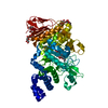
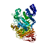
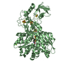
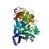
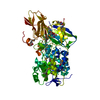
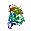
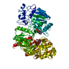
| Title | Crystal Structure of Amylosucrase from Neisseria polysaccharea in Complex with D-glucose | ||||||
 Components Components | Amylosucrase | ||||||
 Keywords Keywords | TRANSFERASE / D-glucose complex | ||||||
| Function / homology |  Function and homology information Function and homology informationamylosucrase / amylosucrase activity / carbohydrate metabolic process / extracellular region Similarity search - Function | ||||||
| Biological species |  Neisseria polysaccharea (bacteria) Neisseria polysaccharea (bacteria) | ||||||
| Method |  X-RAY DIFFRACTION / SYNCHROTRON / Resolution: 1.66 Å X-RAY DIFFRACTION / SYNCHROTRON / Resolution: 1.66 Å | ||||||
 Authors Authors | Mirza, O. / Skov, L.K. / Gajhede, M. | ||||||
 Citation Citation | Journal: Biochemistry / Year: 2001 Title: Crystal structures of amylosucrase from Neisseria polysaccharea in complex with D-glucose and the active site mutant Glu328Gln in complex with the natural substrate sucrose. Authors: Mirza, O. / Skov, L.K. / Remaud-Simeon, M. / Potocki de Montalk, G. / Albenne, C. / Monsan, P. / Gajhede, M. | ||||||
| History |
| ||||||
| Remark 999 | SEQUENCE THE DISCREPANCY BETWEEN RESIDUE ASP 537 AND THE GENBANK SEQUENCE DATABASE REFERENCE, ...SEQUENCE THE DISCREPANCY BETWEEN RESIDUE ASP 537 AND THE GENBANK SEQUENCE DATABASE REFERENCE, ACCESSION 4107260, RESIDUE GLY 545 IS DUE TO A PCR ERROR. |
- Structure visualization
| Structure viewer | Molecule: MolmilJmol/JSmol |
|---|
- Downloads & links
-Download
| PDBx/mmCIF format | 1jg9.cif.gz | 157.1 KB | Display | PDBx/mmCIF format |
|---|---|---|---|---|
| PDB format | pdb1jg9.ent.gz | 121.5 KB | Display | PDB format |
| PDBx/mmJSON format | 1jg9.json.gz | Tree view | PDBx/mmJSON format | |
| Others |  Other downloads Other downloads |
-Validation report
| Arichive directory | https://data.pdbj.org/pub/pdb/validation_reports/jg/1jg9ftp://data.pdbj.org/pub/pdb/validation_reports/jg/1jg9 | HTTPS FTP |
|---|
-Related structure data


-Links
 PDBj
PDBj


- Assembly
| Deposited unit | 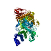
| ||||||||
|---|---|---|---|---|---|---|---|---|---|
| 1 |
| ||||||||
| Unit cell |
|
-Components
| #1: Protein | Mass: 71564.078 Da / Num. of mol.: 1 Source method: isolated from a genetically manipulated source Source: (gene. exp.) Neisseria polysaccharea (bacteria) / Production host: | ||
|---|---|---|---|
| #2: Sugar |   Type: D-saccharide, alpha linking / Mass: 180.156 Da / Num. of mol.: 3 Type: D-saccharide, alpha linking / Mass: 180.156 Da / Num. of mol.: 3Source method: isolated from a genetically manipulated source Formula: C6H12O6 #3: Water | ChemComp-HOH / |  Mass: 18.015 Da / Num. of mol.: 890 / Source method: isolated from a natural source / Formula: H2O Mass: 18.015 Da / Num. of mol.: 890 / Source method: isolated from a natural source / Formula: H2O |
-Experimental details
-Experiment
| Experiment | Method: X-RAY DIFFRACTION / Number of used crystals: 1 |
|---|
- Sample preparation
| Crystal | Density Matthews: 2.4 Å3/Da / Density % sol: 48.79 % | ||||||||||||||||||||||||||||||||||||||||||||||||
|---|---|---|---|---|---|---|---|---|---|---|---|---|---|---|---|---|---|---|---|---|---|---|---|---|---|---|---|---|---|---|---|---|---|---|---|---|---|---|---|---|---|---|---|---|---|---|---|---|---|
| Crystal grow | *PLUS Temperature: 4 ℃ / pH: 7 / Method: vapor diffusion, hanging drop | ||||||||||||||||||||||||||||||||||||||||||||||||
| Components of the solutions | *PLUS
|
-Data collection
| Diffraction | Mean temperature: 120 K |
|---|---|
| Diffraction source | Source: SYNCHROTRON / Site: ESRF  / Beamline: ID14-1 / Wavelength: 0.933 Å / Beamline: ID14-1 / Wavelength: 0.933 Å |
| Detector | Type: MARRESEARCH / Detector: CCD / Date: Dec 10, 1999 |
| Radiation | Protocol: SINGLE WAVELENGTH / Monochromatic (M) / Laue (L): M / Scattering type: x-ray |
| Radiation wavelength | Wavelength: 0.933 Å / Relative weight: 1 |
| Reflection | Resolution: 1.66→13 Å / Num. obs: 619347 / % possible obs: 99.3 % / Redundancy: 7 % / Biso Wilson estimate: 15 Å2 / Rmerge(I) obs: 0.069 / Net I/σ(I): 9 |
| Reflection shell | Resolution: 1.66→1.72 Å / Redundancy: 4 % / Rmerge(I) obs: 0.076 / Mean I/σ(I) obs: 2.5 / Num. unique all: 8012 / % possible all: 99.4 |
| Reflection | *PLUS Lowest resolution: 13 Å / Num. obs: 81373 / Num. measured all: 619347 |
| Reflection shell | *PLUS % possible obs: 99.4 % / Mean I/σ(I) obs: 0.338 |
- Processing
| Software |
| ||||||||||||||||||||||||||||||||||||||||||||
|---|---|---|---|---|---|---|---|---|---|---|---|---|---|---|---|---|---|---|---|---|---|---|---|---|---|---|---|---|---|---|---|---|---|---|---|---|---|---|---|---|---|---|---|---|---|
| Refinement | Resolution: 1.66→12.98 Å / Rfactor Rfree error: 0.003 / Data cutoff high absF: 536057.59 / Data cutoff low absF: 0 / Isotropic thermal model: RESTRAINED / Cross valid method: THROUGHOUT / σ(F): 0
| ||||||||||||||||||||||||||||||||||||||||||||
| Solvent computation | Solvent model: FLAT MODEL / Bsol: 44.8357 Å2 / ksol: 0.396523 e/Å3 | ||||||||||||||||||||||||||||||||||||||||||||
| Displacement parameters | Biso mean: 16.2 Å2
| ||||||||||||||||||||||||||||||||||||||||||||
| Refine analyze |
| ||||||||||||||||||||||||||||||||||||||||||||
| Refinement step | Cycle: LAST / Resolution: 1.66→12.98 Å
| ||||||||||||||||||||||||||||||||||||||||||||
| Refine LS restraints |
| ||||||||||||||||||||||||||||||||||||||||||||
| LS refinement shell | Resolution: 1.66→1.76 Å / Rfactor Rfree error: 0.01 / Total num. of bins used: 6
| ||||||||||||||||||||||||||||||||||||||||||||
| Xplor file |
| ||||||||||||||||||||||||||||||||||||||||||||
| Software | *PLUS Name: CNS / Version: 1 / Classification: refinement | ||||||||||||||||||||||||||||||||||||||||||||
| Refinement | *PLUS σ(F): 0 / % reflection Rfree: 5 % / Rfactor obs: 0.185 | ||||||||||||||||||||||||||||||||||||||||||||
| Solvent computation | *PLUS | ||||||||||||||||||||||||||||||||||||||||||||
| Displacement parameters | *PLUS Biso mean: 16.2 Å2 | ||||||||||||||||||||||||||||||||||||||||||||
| Refine LS restraints | *PLUS
| ||||||||||||||||||||||||||||||||||||||||||||
| LS refinement shell | *PLUS Rfactor Rfree: 0.233 / % reflection Rfree: 4.8 % / Rfactor Rwork: 0.218 |
