 Movie
Movie Controller


+ Open data

- Basic information
| Entry | Database: PDB / ID: 1j1j | ||||||
|---|---|---|---|---|---|---|---|
| Title | Crystal Structure of human Translin | ||||||
 Components Components | Translin | ||||||
 Keywords Keywords | DNA BINDING PROTEIN / Testis/brain RNA binding protein / ssDNA binding protein / RNA binding protein | ||||||
| Function / homology |  Function and homology information Function and homology informationendoribonuclease complex / Small interfering RNA (siRNA) biogenesis / regulatory ncRNA-mediated post-transcriptional gene silencing / siRNA processing / male germ cell nucleus / single-stranded DNA binding / endonuclease activity / sequence-specific DNA binding / Hydrolases; Acting on ester bonds / mRNA binding ...endoribonuclease complex / Small interfering RNA (siRNA) biogenesis / regulatory ncRNA-mediated post-transcriptional gene silencing / siRNA processing / male germ cell nucleus / single-stranded DNA binding / endonuclease activity / sequence-specific DNA binding / Hydrolases; Acting on ester bonds / mRNA binding / endoplasmic reticulum / DNA binding / RNA binding / nucleoplasm / identical protein binding / nucleus / cytoplasm / cytosol Similarity search - Function | ||||||
| Biological species |  Homo sapiens (human) Homo sapiens (human) | ||||||
| Method |  X-RAY DIFFRACTION / SYNCHROTRON / MAD / Resolution: 2.2 Å X-RAY DIFFRACTION / SYNCHROTRON / MAD / Resolution: 2.2 Å | ||||||
 Authors Authors | Sugiura, I. / Sasaki, C. / Hasegawa, T. / Kohno, T. / Sugio, S. / Moriyama, H. / Kasai, M. / Matsuzaki, T. | ||||||
 Citation Citation | Journal: Acta Crystallogr.,Sect.D / Year: 2004 Title: Structure of human translin at 2.2 A resolution. Authors: Sugiura, I. / Sasaki, C. / Hasegawa, T. / Kohno, T. / Sugio, S. / Moriyama, H. / Kasai, M. / Matsuzaki, T. | ||||||
| History |
|
- Structure visualization
| Structure viewer | Molecule: MolmilJmol/JSmol |
|---|
- Downloads & links
-Download
| PDBx/mmCIF format | 1j1j.cif.gz | 189.1 KB | Display | PDBx/mmCIF format |
|---|---|---|---|---|
| PDB format | pdb1j1j.ent.gz | 153.1 KB | Display | PDB format |
| PDBx/mmJSON format | 1j1j.json.gz | Tree view | PDBx/mmJSON format | |
| Others |  Other downloads Other downloads |
-Validation report
| Arichive directory | https://data.pdbj.org/pub/pdb/validation_reports/j1/1j1jftp://data.pdbj.org/pub/pdb/validation_reports/j1/1j1j | HTTPS FTP |
|---|
-Related structure data
| Related structure data | |
|---|---|
| Similar structure data |


-Links
 PDBj
PDBj
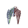
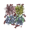
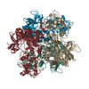
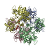
- Assembly
| Deposited unit | 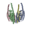
| ||||||||
|---|---|---|---|---|---|---|---|---|---|
| 1 |
| ||||||||
| 2 | 
| ||||||||
| 3 | 
| ||||||||
| 4 | 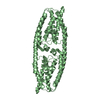
| ||||||||
| 5 | 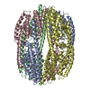
| ||||||||
| Unit cell |
|
-Components
| #1: Protein | Mass: 27625.498 Da / Num. of mol.: 4 Source method: isolated from a genetically manipulated source Source: (gene. exp.) Homo sapiens (human) / Plasmid: pQE9 / Production host:  #2: Water | ChemComp-HOH / |  Mass: 18.015 Da / Num. of mol.: 414 / Source method: isolated from a natural source / Formula: H2O Mass: 18.015 Da / Num. of mol.: 414 / Source method: isolated from a natural source / Formula: H2O |
|---|
-Experimental details
-Experiment
| Experiment | Method: X-RAY DIFFRACTION / Number of used crystals: 1 |
|---|
- Sample preparation
| Crystal | Density Matthews: 2.8 Å3/Da / Density % sol: 55.76 % | |||||||||||||||||||||||||||||||||||||||||||||||||
|---|---|---|---|---|---|---|---|---|---|---|---|---|---|---|---|---|---|---|---|---|---|---|---|---|---|---|---|---|---|---|---|---|---|---|---|---|---|---|---|---|---|---|---|---|---|---|---|---|---|---|
| Crystal grow | Temperature: 293 K / Method: vapor diffusion, sitting drop / pH: 4.5 Details: Sodium Formate, Sodium Chloride, Acetate, pH 4.5, VAPOR DIFFUSION, SITTING DROP, temperature 293K | |||||||||||||||||||||||||||||||||||||||||||||||||
| Crystal grow | *PLUS Temperature: 293 K / pH: 7 / Method: vapor diffusion, hanging drop | |||||||||||||||||||||||||||||||||||||||||||||||||
| Components of the solutions | *PLUS
|
-Data collection
| Diffraction | Mean temperature: 100 K |
|---|---|
| Diffraction source | Source: SYNCHROTRON / Site: SPring-8  / Beamline: BL24XU / Wavelength: 0.836 Å / Beamline: BL24XU / Wavelength: 0.836 Å |
| Detector | Type: RIGAKU RAXIS IV / Detector: IMAGE PLATE / Date: Mar 5, 2000 |
| Radiation | Monochromator: Si III CHANNEL / Protocol: SINGLE WAVELENGTH / Monochromatic (M) / Laue (L): M / Scattering type: x-ray |
| Radiation wavelength | Wavelength: 0.836 Å / Relative weight: 1 |
| Reflection | Resolution: 2.2→40 Å / Num. obs: 57661 / % possible obs: 97.3 % / Observed criterion σ(I): 3 / Redundancy: 5.1 % |
| Reflection shell | Resolution: 2.2→2.32 Å / Redundancy: 4.7 % / Mean I/σ(I) obs: 1.8 / Num. unique all: 8420 / Rsym value: 0.37 / % possible all: 97.3 |
| Reflection | *PLUS Highest resolution: 2.2 Å / Lowest resolution: 15 Å / Num. obs: 57689 / % possible obs: 97.3 % / Rmerge(I) obs: 0.087 |
| Reflection shell | *PLUS Highest resolution: 2.2 Å / Lowest resolution: 2.32 Å / % possible obs: 97.3 % / Rmerge(I) obs: 0.414 / Mean I/σ(I) obs: 3.5 |
- Processing
| Software |
| ||||||||||||||||||||||||
|---|---|---|---|---|---|---|---|---|---|---|---|---|---|---|---|---|---|---|---|---|---|---|---|---|---|
| Refinement | Method to determine structure: MAD / Resolution: 2.2→15 Å / σ(F): 0 / Stereochemistry target values: Engh & Huber
| ||||||||||||||||||||||||
| Refinement step | Cycle: LAST / Resolution: 2.2→15 Å
| ||||||||||||||||||||||||
| Refinement | *PLUS Lowest resolution: 15 Å | ||||||||||||||||||||||||
| Solvent computation | *PLUS | ||||||||||||||||||||||||
| Displacement parameters | *PLUS | ||||||||||||||||||||||||
| Refine LS restraints | *PLUS
|
