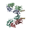
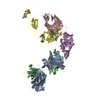
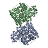
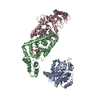
[English] 日本語
Yorodumi
- PDB-1ivo: Crystal Structure of the Complex of Human Epidermal Growth Factor... -

+
Open data


ID or keywords:

Loading...

-
Basic information

Entry
Database: PDB / ID: 1ivo
TitleCrystal Structure of the Complex of Human Epidermal Growth Factor and Receptor Extracellular Domains.
Components
  • Epidermal Growth Factor Receptor
  • Epidermal growth factor
KeywordsTRANSFERASE/SIGNALING PROTEIN / Transmembrane / Glycoprotein / Receptor / RIKEN Structural Genomics/Proteomics Initiative / RSGI / Structural Genomics / TRANSFERASE-SIGNALING PROTEIN COMPLEX
Function / homology
Function and homology information


Inhibition of Signaling by Overexpressed EGFR / EGFR interacts with phospholipase C-gamma / negative regulation of secretion / negative regulation of cholesterol efflux / positive regulation of hyaluronan biosynthetic process / PLCG1 events in ERBB2 signaling / positive regulation of cerebellar granule cell precursor proliferation / cerebellar granule cell precursor proliferation / ERBB2-EGFR signaling pathway / positive regulation of epithelial tube formation ...Inhibition of Signaling by Overexpressed EGFR / EGFR interacts with phospholipase C-gamma / negative regulation of secretion / negative regulation of cholesterol efflux / positive regulation of hyaluronan biosynthetic process / PLCG1 events in ERBB2 signaling / positive regulation of cerebellar granule cell precursor proliferation / cerebellar granule cell precursor proliferation / ERBB2-EGFR signaling pathway / positive regulation of epithelial tube formation / ERBB2 Activates PTK6 Signaling / regulation of calcium ion import / positive regulation of protein localization to early endosome / regulation of protein localization to cell surface / Signaling by EGFR / transmembrane receptor protein tyrosine kinase activator activity / Signaling by ERBB4 / positive regulation of ubiquitin-dependent protein catabolic process / ERBB2 Regulates Cell Motility / Differentiation of keratinocytes in interfollicular epidermis in mammalian skin / PI3K events in ERBB2 signaling / regulation of receptor signaling pathway via JAK-STAT / Estrogen-dependent nuclear events downstream of ESR-membrane signaling / NFE2L2 regulating tumorigenic genes / epidermal growth factor receptor binding / branching morphogenesis of an epithelial tube / positive regulation of DNA binding / GAB1 signalosome / positive regulation of phosphorylation / positive regulation of receptor internalization / Signaling by ERBB2 / GRB2 events in EGFR signaling / SHC1 events in EGFR signaling / Complex I biogenesis / mammary gland alveolus development / GRB2 events in ERBB2 signaling / Respiratory electron transport / SHC1 events in ERBB2 signaling / epithelial cell proliferation / positive regulation of endothelial cell proliferation / ERK1 and ERK2 cascade / NOTCH3 Activation and Transmission of Signal to the Nucleus / positive regulation of endothelial cell migration / positive regulation of mitotic nuclear division / EGFR downregulation / Signaling by ERBB2 TMD/JMD mutants / platelet alpha granule lumen / guanyl-nucleotide exchange factor activity / : / positive regulation of peptidyl-threonine phosphorylation / Constitutive Signaling by EGFRvIII / clathrin-coated endocytic vesicle membrane / mitochondrial electron transport, NADH to ubiquinone / Signaling by ERBB2 ECD mutants / proton motive force-driven mitochondrial ATP synthesis / Signaling by ERBB2 KD Mutants / epidermal growth factor receptor signaling pathway / NADH dehydrogenase (ubiquinone) activity / Downregulation of ERBB2 signaling / growth factor activity / aerobic respiration / Constitutive Signaling by Aberrant PI3K in Cancer / positive regulation of canonical Wnt signaling pathway / Cargo recognition for clathrin-mediated endocytosis / positive regulation of peptidyl-tyrosine phosphorylation / Constitutive Signaling by Ligand-Responsive EGFR Cancer Variants / PIP3 activates AKT signaling / Clathrin-mediated endocytosis / Platelet degranulation / PI5P, PP2A and IER3 Regulate PI3K/AKT Signaling / RAF/MAP kinase cascade / angiogenesis / Extra-nuclear estrogen signaling / positive regulation of MAPK cascade / positive regulation of phosphatidylinositol 3-kinase/protein kinase B signal transduction / receptor ligand activity / electron transfer activity / mitochondrial inner membrane / positive regulation of cell migration / lysosomal membrane / positive regulation of cell population proliferation / calcium ion binding / positive regulation of gene expression / positive regulation of DNA-templated transcription / extracellular space / extracellular exosome / extracellular region / plasma membrane
Similarity search - Function
Pro-epidermal growth factor / 24 nucleotide stem-loop, u2 snrnp hairpin iv. U2 a'; Chain A / Receptor L-domain / : / Hormone Receptor, Insulin-like Growth Factor Receptor 1; Chain A domain 2 / Hormone Receptor, Insulin-like Growth Factor Receptor 1; Chain A, domain 2 / Growth factor receptor domain IV / Low-density lipoprotein receptor repeat class B / LDL-receptor class B (LDLRB) repeat profile. / LDLR class B repeat ...Pro-epidermal growth factor / 24 nucleotide stem-loop, u2 snrnp hairpin iv. U2 a'; Chain A / Receptor L-domain / : / Hormone Receptor, Insulin-like Growth Factor Receptor 1; Chain A domain 2 / Hormone Receptor, Insulin-like Growth Factor Receptor 1; Chain A, domain 2 / Growth factor receptor domain IV / Low-density lipoprotein receptor repeat class B / LDL-receptor class B (LDLRB) repeat profile. / LDLR class B repeat / Low-density lipoprotein-receptor YWTD domain / : / Calcium-binding EGF domain / Furin-like cysteine rich region / Receptor L domain / Alpha-Beta Horseshoe / Six-bladed beta-propeller, TolB-like / Laminin / Coagulation Factor Xa inhibitory site / Laminin / EGF-like domain / NADH dehydrogenase [ubiquinone] iron-sulfur protein 6, mitochondrial / EGF-type aspartate/asparagine hydroxylation site / EGF-like calcium-binding, conserved site / Zinc finger, CHCC-type / Zinc-finger domain / Calcium-binding EGF-like domain signature. / Aspartic acid and asparagine hydroxylation site. / EGF-like calcium-binding domain / Calcium-binding EGF-like domain / Growth factor receptor cysteine-rich domain superfamily / Epidermal growth factor-like domain. / EGF-like domain profile. / EGF-like domain signature 2. / EGF-like domain signature 1. / EGF-like domain / Ribbon / Mainly Beta / Alpha Beta
Similarity search - Domain/homology
NADH dehydrogenase [ubiquinone] iron-sulfur protein 6, mitochondrial / Pro-epidermal growth factor
Similarity search - Component
Biological speciesHomo sapiens (human)
MethodX-RAY DIFFRACTION / SYNCHROTRON / MAD / Resolution: 3.3 Å
AuthorsOgiso, H. / Ishitani, R. / Nureki, O. / Fukai, S. / Yamanaka, M. / Kim, J.H. / Saito, K. / Shirouzu, M. / Yokoyama, S. / RIKEN Structural Genomics/Proteomics Initiative (RSGI)
CitationJournal: Cell(Cambridge,Mass.) / Year: 2002
Title: Crystal Structure of the Complex of Human Epidermal Growth Factor and Receptor Extracellular Domains.
Authors: Ogiso, H. / Ishitani, R. / Nureki, O. / Fukai, S. / Yamanaka, M. / Kim, J.H. / Saito, K. / Inoue, M. / Shirouzu, M. / Yokoyama, S.
History
DepositionMar 28, 2002Deposition site: PDBJ / Processing site: PDBJ
Revision 1.0Oct 16, 2002Provider: repository / Type: Initial release
Revision 1.1Apr 27, 2008Group: Version format compliance
Revision 1.2Jul 13, 2011Group: Non-polymer description / Version format compliance
Revision 2.0Jul 29, 2020Group: Advisory / Atomic model ...Advisory / Atomic model / Data collection / Derived calculations / Structure summary
Category: atom_site / chem_comp ...atom_site / chem_comp / entity / pdbx_branch_scheme / pdbx_chem_comp_identifier / pdbx_entity_branch / pdbx_entity_branch_descriptor / pdbx_entity_branch_link / pdbx_entity_branch_list / pdbx_entity_nonpoly / pdbx_nonpoly_scheme / pdbx_struct_assembly_gen / pdbx_unobs_or_zero_occ_residues / struct_asym / struct_conn / struct_site / struct_site_gen
Item: _atom_site.B_iso_or_equiv / _atom_site.Cartn_x ..._atom_site.B_iso_or_equiv / _atom_site.Cartn_x / _atom_site.Cartn_y / _atom_site.Cartn_z / _atom_site.auth_asym_id / _atom_site.auth_seq_id / _atom_site.label_asym_id / _atom_site.label_entity_id / _chem_comp.name / _chem_comp.type / _pdbx_entity_nonpoly.entity_id / _pdbx_entity_nonpoly.name / _pdbx_struct_assembly_gen.asym_id_list / _struct_conn.pdbx_leaving_atom_flag / _struct_conn.pdbx_role / _struct_conn.ptnr1_auth_asym_id / _struct_conn.ptnr1_auth_seq_id / _struct_conn.ptnr1_label_asym_id / _struct_conn.ptnr2_auth_asym_id / _struct_conn.ptnr2_auth_seq_id / _struct_conn.ptnr2_label_asym_id
Description: Carbohydrate remediation / Provider: repository / Type: Remediation
Revision 2.1Dec 27, 2023Group: Advisory / Data collection ...Advisory / Data collection / Database references / Structure summary
Category: chem_comp / chem_comp_atom ...chem_comp / chem_comp_atom / chem_comp_bond / database_2 / pdbx_unobs_or_zero_occ_residues
Item: _chem_comp.pdbx_synonyms / _database_2.pdbx_DOI / _database_2.pdbx_database_accession

-
Structure visualization

Structure viewerMolecule:
MolmilJmol/JSmol

Downloads & links

-
Assembly

Deposited unit
A: Epidermal Growth Factor Receptor
B: Epidermal Growth Factor Receptor
C: Epidermal growth factor
D: Epidermal growth factor
hetero molecules


Theoretical massNumber of molelcules
Total (without water)152,21313
Polymers150,0194
Non-polymers2,1949
Water1,42379
1


  • Idetical with deposited unit
  • defined by author&software
TypeNameSymmetry operationNumber
identity operation1_555x,y,z1
Buried area11490 Å2
ΔGint-9 kcal/mol
Surface area52390 Å2
MethodPISA
Unit cell
Length a, b, c (Å)220.164, 220.164, 113.121
Angle α, β, γ (deg.)90.00, 90.00, 120.00
Int Tables number152
Space group name H-MP3121

-
Components

#1: Protein Epidermal Growth Factor Receptor


Mass: 68780.328 Da / Num. of mol.: 2 / Fragment: extracellular domains I, II, II and IV
Source method: isolated from a genetically manipulated source
Source: (gene. exp.) Homo sapiens (human) / Plasmid: pcDNA-sEGFR / Production host: Cricetulus griseus (Chinese hamster) / Strain (production host): LEC8 / References: UniProt: P00533, EC: 2.7.1.112
#2: Protein Epidermal growth factor / EGF


Mass: 6229.027 Da / Num. of mol.: 2 / Fragment: RESIDUES 1-53
Source method: isolated from a genetically manipulated source
Source: (gene. exp.) Homo sapiens (human) / Production host: Escherichia coli (E. coli) / References: UniProt: P01133
#3: Polysaccharide 2-acetamido-2-deoxy-beta-D-glucopyranose-(1-4)-2-acetamido-2-deoxy-beta-D-glucopyranose


Type: oligosaccharide / Mass: 424.401 Da / Num. of mol.: 1
Source method: isolated from a genetically manipulated source
DescriptorTypeProgram
DGlcpNAcb1-4DGlcpNAcb1-Glycam Condensed SequenceGMML 1.0
WURCS=2.0/1,2,1/[a2122h-1b_1-5_2*NCC/3=O]/1-1/a4-b1WURCSPDB2Glycan 1.1.0
[]{[(4+1)][b-D-GlcpNAc]{[(4+1)][b-D-GlcpNAc]{}}}LINUCSPDB-CARE
#4: Sugar
ChemComp-NAG / 2-acetamido-2-deoxy-beta-D-glucopyranose / N-acetyl-beta-D-glucosamine / 2-acetamido-2-deoxy-beta-D-glucose / 2-acetamido-2-deoxy-D-glucose / 2-acetamido-2-deoxy-glucose / N-ACETYL-D-GLUCOSAMINE


Type: D-saccharide, beta linking / Mass: 221.208 Da / Num. of mol.: 8
Source method: isolated from a genetically manipulated source
Formula: C8H15NO6
IdentifierTypeProgram
DGlcpNAcbCONDENSED IUPAC CARBOHYDRATE SYMBOLGMML 1.0
N-acetyl-b-D-glucopyranosamineCOMMON NAMEGMML 1.0
b-D-GlcpNAcIUPAC CARBOHYDRATE SYMBOLPDB-CARE 1.0
GlcNAcSNFG CARBOHYDRATE SYMBOLGMML 1.0
#5: Water ChemComp-HOH / water


Mass: 18.015 Da / Num. of mol.: 79 / Source method: isolated from a natural source / Formula: H2O

-
Experimental details

-
Experiment

ExperimentMethod: X-RAY DIFFRACTION / Number of used crystals: 2

-
Sample preparation

CrystalDensity Matthews: 5.28 Å3/Da / Density % sol: 76.69 %
Crystal growTemperature: 293 K / Method: vapor diffusion, hanging drop / pH: 8.4
Details: PEG4000, PEG6000, sodium acetate, sodium chloride, TRIS, pH 8.4, VAPOR DIFFUSION, HANGING DROP, temperature 293K
Crystal grow
*PLUS
Temperature: 20 ℃
Components of the solutions
*PLUS
IDConc.Common nameCrystal-IDSol-IDDetailsChemical formula
115 %PEG40001reservoir
21 %PEG60001reservoir
375 mMTris-HCl1reservoirpH8.4
475 mMsodium acetate1reservoir
5200 mM1reservoirNaCl
610 mg/mlprotein1drop

-
Data collection

Diffraction
IDMean temperature (K)Crystal-ID
11001
21001
Diffraction source
SourceSiteBeamlineIDWavelength (Å)
SYNCHROTRONSPring-8 BL41XU11
SYNCHROTRONSPring-8 BL41XU20.9795, 0.9798, 0.9733, 0.9839
Detector
TypeIDDetectorDetailsDate
MARRESEARCH1CCDmirrors
MARRESEARCH2CCDmirrorsJan 1, 2001
Radiation
IDMonochromatorProtocolMonochromatic (M) / Laue (L)Scattering typeWavelength-ID
1Si(111)SINGLE WAVELENGTHMx-ray1
2Si(111)MADMx-ray1
Radiation wavelength
IDWavelength (Å)Relative weight
111
20.97951
30.97981
40.97331
50.98391
ReflectionResolution: 3.5→50 Å / Num. all: 293584 / Num. obs: 293584 / % possible obs: 99 % / Biso Wilson estimate: 71.3 Å2 / Rmerge(I) obs: 0.075 / Rsym value: 0.075 / Net I/σ(I): 25.6
Reflection shellResolution: 3.5→3.71 Å / Rmerge(I) obs: 0.27 / Mean I/σ(I) obs: 5.1 / Rsym value: 0.27 / % possible all: 98.6
Reflection
*PLUS
Highest resolution: 3.3 Å / Lowest resolution: 50 Å / Num. obs: 46667 / % possible obs: 98.2 % / Num. measured all: 320883 / Rmerge(I) obs: 0.078
Reflection shell
*PLUS
% possible obs: 92.6 % / Rmerge(I) obs: 0.358 / Mean I/σ(I) obs: 2.9

-
Processing

Software
NameVersionClassification
DENZOdata reduction
SCALEPACKdata scaling
MLPHAREphasing
CNS1refinement
RefinementMethod to determine structure: MAD / Resolution: 3.3→10 Å / Rfactor Rfree error: 0.007 / Isotropic thermal model: RESTRAINED / Cross valid method: THROUGHOUT / σ(F): 0 / Stereochemistry target values: Engh & Huber
RfactorNum. reflection% reflectionSelection details
Rfree0.326 2239 5 %RANDOM
Rwork0.255 ---
obs0.258 44854 98 %-
Solvent computationSolvent model: FLAT MODEL / Bsol: 26.4363 Å2 / ksol: 0.166501 e/Å3
Displacement parametersBiso mean: 82.5 Å2
Baniso -1Baniso -2Baniso -3
1--9.51 Å215.74 Å20 Å2
2---9.51 Å20 Å2
3---19.03 Å2
Refine analyze
FreeObs
Luzzati coordinate error0.54 Å0.39 Å
Luzzati d res low-5 Å
Luzzati sigma a0.75 Å0.5 Å
Refinement stepCycle: LAST / Resolution: 3.3→10 Å
ProteinNucleic acidLigandSolventTotal
Num. atoms8673 0 140 79 8892
Refine LS restraints
Refine-IDTypeDev ideal
X-RAY DIFFRACTIONc_bond_d0.006
X-RAY DIFFRACTIONc_bond_d_na
X-RAY DIFFRACTIONc_bond_d_prot
X-RAY DIFFRACTIONc_angle_d
X-RAY DIFFRACTIONc_angle_d_na
X-RAY DIFFRACTIONc_angle_d_prot
X-RAY DIFFRACTIONc_angle_deg1.4
X-RAY DIFFRACTIONc_angle_deg_na
X-RAY DIFFRACTIONc_angle_deg_prot
X-RAY DIFFRACTIONc_dihedral_angle_d25.1
X-RAY DIFFRACTIONc_dihedral_angle_d_na
X-RAY DIFFRACTIONc_dihedral_angle_d_prot
X-RAY DIFFRACTIONc_improper_angle_d0.78
X-RAY DIFFRACTIONc_improper_angle_d_na
X-RAY DIFFRACTIONc_improper_angle_d_prot
X-RAY DIFFRACTIONc_mcbond_it
X-RAY DIFFRACTIONc_mcangle_it
X-RAY DIFFRACTIONc_scbond_it
X-RAY DIFFRACTIONc_scangle_it
Refine LS restraints NCSNCS model details: CONSTR
LS refinement shellResolution: 3.3→3.5 Å / Rfactor Rfree error: 0.02 / Total num. of bins used: 6
RfactorNum. reflection% reflection
Rfree0.37 360 5.2 %
Rwork0.31 6616 -
obs-6103 91.7 %
Xplor file
Refine-IDSerial noParam fileTopol file
X-RAY DIFFRACTION1PROTEIN_REP.PARAMPROTEIN.TOP
X-RAY DIFFRACTION2WATER_REP.PARAMWATER.TOP
X-RAY DIFFRACTION3CARBOHYDRATE.PARAMCARBOHYDRATE.TOP
Refinement
*PLUS
Highest resolution: 3.3 Å / Lowest resolution: 10 Å / % reflection Rfree: 5 %
Solvent computation
*PLUS
Displacement parameters
*PLUS
Refine LS restraints
*PLUS
Refine-IDTypeDev ideal
X-RAY DIFFRACTIONc_angle_deg1.39
X-RAY DIFFRACTIONc_dihedral_angle_d
X-RAY DIFFRACTIONc_dihedral_angle_deg25.1
X-RAY DIFFRACTIONc_improper_angle_d
X-RAY DIFFRACTIONc_improper_angle_deg0.781

+
About Yorodumi

-
News

-
Feb 9, 2022. New format data for meta-information of EMDB entries

New format data for meta-information of EMDB entries

  • Version 3 of the EMDB header file is now the official format.
  • The previous official version 1.9 will be removed from the archive.

Related info.:EMDB header

External links:wwPDB to switch to version 3 of the EMDB data model

-
Aug 12, 2020. Covid-19 info

Covid-19 info

URL: https://pdbjlvh1.pdbj.org/emnavi/covid19.php

New page: Covid-19 featured information page in EM Navigator.

Related info.:Covid-19 info / Mar 5, 2020. Novel coronavirus structure data

+
Mar 5, 2020. Novel coronavirus structure data

Novel coronavirus structure data

Related info.:Yorodumi Speices / Aug 12, 2020. Covid-19 info

External links:COVID-19 featured content - PDBj / Molecule of the Month (242):Coronavirus Proteases

+
Jan 31, 2019. EMDB accession codes are about to change! (news from PDBe EMDB page)

EMDB accession codes are about to change! (news from PDBe EMDB page)

  • The allocation of 4 digits for EMDB accession codes will soon come to an end. Whilst these codes will remain in use, new EMDB accession codes will include an additional digit and will expand incrementally as the available range of codes is exhausted. The current 4-digit format prefixed with “EMD-” (i.e. EMD-XXXX) will advance to a 5-digit format (i.e. EMD-XXXXX), and so on. It is currently estimated that the 4-digit codes will be depleted around Spring 2019, at which point the 5-digit format will come into force.
  • The EM Navigator/Yorodumi systems omit the EMD- prefix.

Related info.:Q: What is EMD? / ID/Accession-code notation in Yorodumi/EM Navigator

External links:EMDB Accession Codes are Changing Soon! / Contact to PDBj

+
Jul 12, 2017. Major update of PDB

Major update of PDB

  • wwPDB released updated PDB data conforming to the new PDBx/mmCIF dictionary.
  • This is a major update changing the version number from 4 to 5, and with Remediation, in which all the entries are updated.
  • In this update, many items about electron microscopy experimental information are reorganized (e.g. em_software).
  • Now, EM Navigator and Yorodumi are based on the updated data.

External links:wwPDB Remediation / Enriched Model Files Conforming to OneDep Data Standards Now Available in the PDB FTP Archive

-
Yorodumi

Thousand views of thousand structures

  • Yorodumi is a browser for structure data from EMDB, PDB, SASBDB, etc.
  • This page is also the successor to EM Navigator detail page, and also detail information page/front-end page for Omokage search.
  • The word "yorodu" (or yorozu) is an old Japanese word meaning "ten thousand". "mi" (miru) is to see.

Related info.:EMDB / PDB / SASBDB / Comparison of 3 databanks / Yorodumi Search / Aug 31, 2016. New EM Navigator & Yorodumi / Yorodumi Papers / Jmol/JSmol / Function and homology information / Changes in new EM Navigator and Yorodumi

Read more