 Movie
Movie Controller


+ Open data

- Basic information
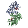
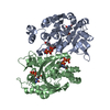
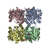
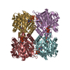
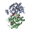
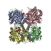
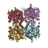
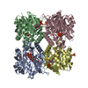
| Entry | Database: PDB / ID: 1iin | ||||||
|---|---|---|---|---|---|---|---|
| Title | thymidylyltransferase complexed with UDP-glucose | ||||||
 Components Components | glucose-1-phosphate thymidylyltransferase | ||||||
 Keywords Keywords | TRANSFERASE | ||||||
| Function / homology |  Function and homology information Function and homology informationglucose-1-phosphate thymidylyltransferase / glucose-1-phosphate thymidylyltransferase activity / O antigen biosynthetic process / dTDP-rhamnose biosynthetic process / polysaccharide biosynthetic process / magnesium ion binding / metal ion binding Similarity search - Function | ||||||
| Biological species |  Salmonella enterica (bacteria) Salmonella enterica (bacteria) | ||||||
| Method |  X-RAY DIFFRACTION / SYNCHROTRON / Resolution: 2.1 Å X-RAY DIFFRACTION / SYNCHROTRON / Resolution: 2.1 Å | ||||||
 Authors Authors | Barton, W.A. / Lesniak, J. / Biggins, J.B. / Jeffrey, P.D. / Jiang, J. / Rajashankar, K.R. / Thorson, J.S. / Nikolov, D.B. | ||||||
 Citation Citation | Journal: Nat.Struct.Biol. / Year: 2001 Title: Structure, mechanism and engineering of a nucleotidylyltransferase as a first step toward glycorandomization. Authors: Barton, W.A. / Lesniak, J. / Biggins, J.B. / Jeffrey, P.D. / Jiang, J. / Rajashankar, K.R. / Thorson, J.S. / Nikolov, D.B. | ||||||
| History |
|
- Structure visualization
| Structure viewer | Molecule: MolmilJmol/JSmol |
|---|
- Downloads & links
-Download
| PDBx/mmCIF format | 1iin.cif.gz | 250.7 KB | Display | PDBx/mmCIF format |
|---|---|---|---|---|
| PDB format | pdb1iin.ent.gz | 202 KB | Display | PDB format |
| PDBx/mmJSON format | 1iin.json.gz | Tree view | PDBx/mmJSON format | |
| Others |  Other downloads Other downloads |
-Validation report
| Arichive directory | https://data.pdbj.org/pub/pdb/validation_reports/ii/1iinftp://data.pdbj.org/pub/pdb/validation_reports/ii/1iin | HTTPS FTP |
|---|
-Related structure data


-Links
 PDBj
PDBj

- Assembly
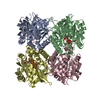
| Deposited unit | 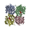
| ||||||||
|---|---|---|---|---|---|---|---|---|---|
| 1 |
| ||||||||
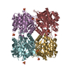
| Unit cell |
|
-Components
| #1: Protein | Mass: 32485.201 Da / Num. of mol.: 4 Source method: isolated from a genetically manipulated source Source: (gene. exp.) Salmonella enterica (bacteria) / Strain: LT2 / Plasmid: pET22 / Production host: References: UniProt: Q9F7K6, UniProt: P26393*PLUS, glucose-1-phosphate thymidylyltransferase #2: Chemical | ChemComp-UPG /   Mass: 566.302 Da / Num. of mol.: 4 / Source method: obtained synthetically / Formula: C15H24N2O17P2 Mass: 566.302 Da / Num. of mol.: 4 / Source method: obtained synthetically / Formula: C15H24N2O17P2#3: Water | ChemComp-HOH / |  Mass: 18.015 Da / Num. of mol.: 762 / Source method: isolated from a natural source / Formula: H2O Mass: 18.015 Da / Num. of mol.: 762 / Source method: isolated from a natural source / Formula: H2O |
|---|
-Experimental details
-Experiment
| Experiment | Method: X-RAY DIFFRACTION / Number of used crystals: 1 |
|---|
- Sample preparation
| Crystal | Density Matthews: 2.65 Å3/Da / Density % sol: 53.58 % | ||||||||||||||||||||||||||||||||||||||||||
|---|---|---|---|---|---|---|---|---|---|---|---|---|---|---|---|---|---|---|---|---|---|---|---|---|---|---|---|---|---|---|---|---|---|---|---|---|---|---|---|---|---|---|---|
| Crystal grow | Temperature: 298 K / Method: vapor diffusion, hanging drop / pH: 7 Details: Ammonium Sulfate, iso-propanol, pH 7, VAPOR DIFFUSION, HANGING DROP, temperature 298K | ||||||||||||||||||||||||||||||||||||||||||
| Crystal grow | *PLUS pH: 7.2 | ||||||||||||||||||||||||||||||||||||||||||
| Components of the solutions | *PLUS
|
-Data collection
| Diffraction | Mean temperature: 130 K |
|---|---|
| Diffraction source | Source: SYNCHROTRON / Site: NSLS  / Beamline: X9B / Wavelength: 0.979 Å / Beamline: X9B / Wavelength: 0.979 Å |
| Detector | Type: CUSTOM-MADE / Detector: CCD |
| Radiation | Protocol: SINGLE WAVELENGTH / Monochromatic (M) / Laue (L): M / Scattering type: x-ray |
| Radiation wavelength | Wavelength: 0.979 Å / Relative weight: 1 |
| Reflection | Resolution: 2→39.83 Å / Num. all: 93944 / Observed criterion σ(F): 0 / Biso Wilson estimate: 9.7 Å2 / Limit h max: 46 / Limit h min: 0 / Limit k max: 56 / Limit k min: 0 / Limit l max: 66 / Limit l min: 0 / Observed criterion F max: 127108.29 / Observed criterion F min: 0.32 |
| Reflection | *PLUS % possible obs: 99.7 % / Redundancy: 7 % / Rmerge(I) obs: 0.075 |
- Processing
| Software |
| ||||||||||||||||||||||||||||||||||||||||||||||||||||||||||||||||||||||||||||||||||||||||||
|---|---|---|---|---|---|---|---|---|---|---|---|---|---|---|---|---|---|---|---|---|---|---|---|---|---|---|---|---|---|---|---|---|---|---|---|---|---|---|---|---|---|---|---|---|---|---|---|---|---|---|---|---|---|---|---|---|---|---|---|---|---|---|---|---|---|---|---|---|---|---|---|---|---|---|---|---|---|---|---|---|---|---|---|---|---|---|---|---|---|---|---|
| Refinement | Resolution: 2.1→39.83 Å / Rfactor Rfree error: 0.002 / Occupancy max: 1 / Occupancy min: 1 / Cross valid method: THROUGHOUT Details: There are three C-terminal residues in the crystal for which the author does not see clear density: LYS 290, GLY 291, LEU 292. There is no density beyond the CB for LYS 154.
| ||||||||||||||||||||||||||||||||||||||||||||||||||||||||||||||||||||||||||||||||||||||||||
| Solvent computation | Solvent model: CNS bulk solvent model used / Bsol: 55.0659 Å2 / ksol: 0.371202 e/Å3 | ||||||||||||||||||||||||||||||||||||||||||||||||||||||||||||||||||||||||||||||||||||||||||
| Displacement parameters | Biso max: 72.5 Å2 / Biso mean: 20.26 Å2 / Biso min: 2.11 Å2
| ||||||||||||||||||||||||||||||||||||||||||||||||||||||||||||||||||||||||||||||||||||||||||
| Refine analyze |
| ||||||||||||||||||||||||||||||||||||||||||||||||||||||||||||||||||||||||||||||||||||||||||
| Refinement step | Cycle: LAST / Resolution: 2.1→39.83 Å
| ||||||||||||||||||||||||||||||||||||||||||||||||||||||||||||||||||||||||||||||||||||||||||
| Refine LS restraints |
| ||||||||||||||||||||||||||||||||||||||||||||||||||||||||||||||||||||||||||||||||||||||||||
| LS refinement shell | Refine-ID: X-RAY DIFFRACTION
| ||||||||||||||||||||||||||||||||||||||||||||||||||||||||||||||||||||||||||||||||||||||||||
| Xplor file |
| ||||||||||||||||||||||||||||||||||||||||||||||||||||||||||||||||||||||||||||||||||||||||||
| Software | *PLUS Name: CNS / Version: 1 / Classification: refinement | ||||||||||||||||||||||||||||||||||||||||||||||||||||||||||||||||||||||||||||||||||||||||||
| Refinement | *PLUS Highest resolution: 2 Å / Lowest resolution: 8 Å / Num. reflection obs: 179416 / Num. reflection Rfree: 2276 / % reflection Rfree: 10 % / Rfactor obs: 0.183 / Rfactor Rfree: 0.223 | ||||||||||||||||||||||||||||||||||||||||||||||||||||||||||||||||||||||||||||||||||||||||||
| Solvent computation | *PLUS | ||||||||||||||||||||||||||||||||||||||||||||||||||||||||||||||||||||||||||||||||||||||||||
| Displacement parameters | *PLUS | ||||||||||||||||||||||||||||||||||||||||||||||||||||||||||||||||||||||||||||||||||||||||||
| Refine LS restraints | *PLUS
| ||||||||||||||||||||||||||||||||||||||||||||||||||||||||||||||||||||||||||||||||||||||||||
| LS refinement shell | *PLUS % reflection Rfree: 9.8 % |
