 Movie
Movie Controller


+ Open data

- Basic information
| Entry | Database: PDB / ID: 1mp5 | ||||||
|---|---|---|---|---|---|---|---|
| Title | Y177F VARIANT OF S. ENTERICA RmlA | ||||||
 Components Components | Y177F VARIANT OF S. ENTERICA RmlA BOUND TO UDP-GLUCOSE | ||||||
 Keywords Keywords | TRANSFERASE | ||||||
| Function / homology |  Function and homology information Function and homology informationglucose-1-phosphate thymidylyltransferase / glucose-1-phosphate thymidylyltransferase activity / O antigen biosynthetic process / dTDP-rhamnose biosynthetic process / polysaccharide biosynthetic process / nucleotide binding / magnesium ion binding / metal ion binding / cytosol Similarity search - Function | ||||||
| Biological species |  Salmonella enterica (bacteria) Salmonella enterica (bacteria) | ||||||
| Method |  X-RAY DIFFRACTION / SYNCHROTRON / MOLECULAR REPLACEMENT / Resolution: 2.75 Å X-RAY DIFFRACTION / SYNCHROTRON / MOLECULAR REPLACEMENT / Resolution: 2.75 Å | ||||||
 Authors Authors | Barton, W.A. / Biggins, J.B. / Jiang, J. / Thorson, J.S. / Nikolov, D.B. | ||||||
 Citation Citation | Journal: Proc.Natl.Acad.Sci.USA / Year: 2002 Title: Expanding pyrimidine diphosphosugar libraries via structure-based nucleotidylyltransferase engineering Authors: Barton, W.A. / Biggins, J.B. / Jiang, J. / Thorson, J.S. / Nikolov, D.B. | ||||||
| History |
| ||||||
| Remark 999 | SEQUENCE Author states the sequence of the deposited model differs from the published sequence, ...SEQUENCE Author states the sequence of the deposited model differs from the published sequence, because there are confirmed natural mutations in the variant of Salmonella used in this entry. |
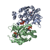
- Structure visualization
| Structure viewer | Molecule: MolmilJmol/JSmol |
|---|
- Downloads & links
-Download
| PDBx/mmCIF format | 1mp5.cif.gz | 228.4 KB | Display | PDBx/mmCIF format |
|---|---|---|---|---|
| PDB format | pdb1mp5.ent.gz | 187 KB | Display | PDB format |
| PDBx/mmJSON format | 1mp5.json.gz | Tree view | PDBx/mmJSON format | |
| Others |  Other downloads Other downloads |
-Validation report
| Arichive directory | https://data.pdbj.org/pub/pdb/validation_reports/mp/1mp5ftp://data.pdbj.org/pub/pdb/validation_reports/mp/1mp5 | HTTPS FTP |
|---|
-Related structure data
| Related structure data |  1mp3C 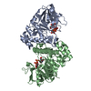 1mp4C  1iinS S: Starting model for refinement C: citing same article ( |
|---|---|
| Similar structure data |


-Links
 PDBj
PDBj

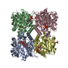
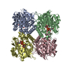
- Assembly
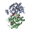
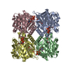
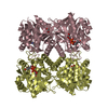
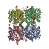
| Deposited unit | 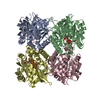
| ||||||||
|---|---|---|---|---|---|---|---|---|---|
| 1 |
| ||||||||
| Unit cell |
|
-Components
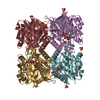
| #1: Protein | Mass: 32411.100 Da / Num. of mol.: 4 / Mutation: Y177F Source method: isolated from a genetically manipulated source Source: (gene. exp.) Salmonella enterica (bacteria) / Production host: References: UniProt: Q9F7G8, UniProt: P26393*PLUS, glucose-1-phosphate thymidylyltransferase #2: Chemical | ChemComp-UPG /   Mass: 566.302 Da / Num. of mol.: 4 / Source method: obtained synthetically / Formula: C15H24N2O17P2 Mass: 566.302 Da / Num. of mol.: 4 / Source method: obtained synthetically / Formula: C15H24N2O17P2 |
|---|
-Experimental details
-Experiment
| Experiment | Method: X-RAY DIFFRACTION / Number of used crystals: 2 |
|---|
- Sample preparation
| Crystal | Density Matthews: 2.58 Å3/Da / Density % sol: 52.31 % | ||||||||||||||||||||||||||||||||||||||||||
|---|---|---|---|---|---|---|---|---|---|---|---|---|---|---|---|---|---|---|---|---|---|---|---|---|---|---|---|---|---|---|---|---|---|---|---|---|---|---|---|---|---|---|---|
| Crystal grow | pH: 7 / Details: pH 7.0, temperature 100K | ||||||||||||||||||||||||||||||||||||||||||
| Crystal grow | *PLUS pH: 7.4 / Method: vapor diffusion, hanging drop | ||||||||||||||||||||||||||||||||||||||||||
| Components of the solutions | *PLUS
|
-Data collection
| Diffraction | Mean temperature: 100 K |
|---|---|
| Diffraction source | Source: SYNCHROTRON / Site: NSLS  / Beamline: X25 / Wavelength: 1.1395 Å / Beamline: X25 / Wavelength: 1.1395 Å |
| Detector | Date: Jun 8, 2001 |
| Radiation | Protocol: SINGLE WAVELENGTH / Monochromatic (M) / Laue (L): M / Scattering type: x-ray |
| Radiation wavelength | Wavelength: 1.1395 Å / Relative weight: 1 |
| Reflection | Resolution: 2.1→50 Å / Num. obs: 43514 / Observed criterion σ(I): 0 |
| Reflection | *PLUS Highest resolution: 2.75 Å / % possible obs: 98.3 % / Redundancy: 5 % / Rmerge(I) obs: 0.061 |
- Processing
| Refinement | Method to determine structure: MOLECULAR REPLACEMENT Starting model: PDB ENTRY 1IIN Resolution: 2.75→50 Å / Cross valid method: THROUGHOUT / σ(F): 0 / Stereochemistry target values: Engh&Huber
| ||||||||||||||||||||
|---|---|---|---|---|---|---|---|---|---|---|---|---|---|---|---|---|---|---|---|---|---|
| Refinement step | Cycle: LAST / Resolution: 2.75→50 Å
| ||||||||||||||||||||
| Refinement | *PLUS Lowest resolution: 8 Å / % reflection Rfree: 10 % / Rfactor Rfree: 0.292 / Rfactor Rwork: 0.229 | ||||||||||||||||||||
| Solvent computation | *PLUS | ||||||||||||||||||||
| Displacement parameters | *PLUS | ||||||||||||||||||||
| Refine LS restraints | *PLUS
|
