 Movie
Movie Controller

[English] 日本語
 Yorodumi
- PDB-1lvw: Crystal structure of glucose-1-phosphate thymidylyltransferase, R... -
+ Open data

- Basic information
| Entry | Database: PDB / ID: 1lvw | ||||||
|---|---|---|---|---|---|---|---|
| Title | Crystal structure of glucose-1-phosphate thymidylyltransferase, RmlA, complex with dTDP | ||||||
 Components Components | glucose-1-phosphate thymidylyltransferase | ||||||
 Keywords Keywords | TRANSFERASE / protein nucleotide complex / nucleotide binding fold / Structural Genomics / PSI / Protein Structure Initiative / Northeast Structural Genomics Consortium / NESG | ||||||
| Function / homology |  Function and homology information Function and homology informationglucose-1-phosphate thymidylyltransferase / glucose-1-phosphate thymidylyltransferase activity / metal ion binding Similarity search - Function | ||||||
| Biological species |   Methanothermobacter thermautotrophicus (archaea) Methanothermobacter thermautotrophicus (archaea) | ||||||
| Method |  X-RAY DIFFRACTION / SYNCHROTRON / MAD / Resolution: 1.7 Å X-RAY DIFFRACTION / SYNCHROTRON / MAD / Resolution: 1.7 Å | ||||||
 Authors Authors | Dong, A. / Christendat, D. / Pai, E.F. / Northeast Structural Genomics Consortium (NESG) | ||||||
 Citation Citation | Journal: To be Published Title: Crystal structure of glucose-1-phosphate thymidylyltransferase, RmlA, complex with dTDP Authors: Dong, A. / Christendat, D. / Pai, E.F. | ||||||
| History |
|
- Structure visualization
| Structure viewer | Molecule: MolmilJmol/JSmol |
|---|
- Downloads & links
-Download
| PDBx/mmCIF format | 1lvw.cif.gz | 266.6 KB | Display | PDBx/mmCIF format |
|---|---|---|---|---|
| PDB format | pdb1lvw.ent.gz | 214.5 KB | Display | PDB format |
| PDBx/mmJSON format | 1lvw.json.gz | Tree view | PDBx/mmJSON format | |
| Others |  Other downloads Other downloads |
-Validation report
| Arichive directory | https://data.pdbj.org/pub/pdb/validation_reports/lv/1lvwftp://data.pdbj.org/pub/pdb/validation_reports/lv/1lvw | HTTPS FTP |
|---|
-Related structure data
| Similar structure data | |
|---|---|
| Other databases |

-Links
 PDBj
PDBj

- Assembly
| Deposited unit | 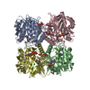
| ||||||||
|---|---|---|---|---|---|---|---|---|---|
| 1 |
| ||||||||
| Unit cell |
| ||||||||
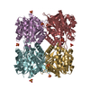
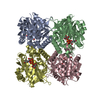
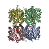
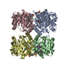
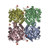
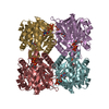
| Details | The biological assembly is a dimer; two dimers in asymmetric unit |
-Components
-Protein , 1 types, 4 molecules ABCD
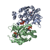
| #1: Protein | Mass: 33366.039 Da / Num. of mol.: 4 Source method: isolated from a genetically manipulated source Source: (gene. exp.) Methanothermobacter thermautotrophicus (archaea)Gene: RMLA / Plasmid: pet15b / Production host:  References: UniProt: O27819, glucose-1-phosphate thymidylyltransferase |
|---|
-Non-polymers , 5 types, 769 molecules 


| #2: Chemical | ChemComp-CL /  Mass: 35.453 Da / Num. of mol.: 5 / Source method: obtained synthetically / Formula: Cl Mass: 35.453 Da / Num. of mol.: 5 / Source method: obtained synthetically / Formula: Cl#3: Chemical |  Mass: 96.063 Da / Num. of mol.: 2 / Source method: obtained synthetically / Formula: SO4 Mass: 96.063 Da / Num. of mol.: 2 / Source method: obtained synthetically / Formula: SO4#4: Chemical | ChemComp-TYD /  Mass: 402.188 Da / Num. of mol.: 8 / Source method: obtained synthetically / Formula: C10H16N2O11P2 Mass: 402.188 Da / Num. of mol.: 8 / Source method: obtained synthetically / Formula: C10H16N2O11P2#5: Chemical | ChemComp-GOL /  Mass: 92.094 Da / Num. of mol.: 11 / Source method: obtained synthetically / Formula: C3H8O3 Mass: 92.094 Da / Num. of mol.: 11 / Source method: obtained synthetically / Formula: C3H8O3#6: Water | ChemComp-HOH / | Mass: 18.015 Da / Num. of mol.: 743 / Source method: isolated from a natural source / Formula: H2O |
|---|
-Experimental details
-Experiment
| Experiment | Method: X-RAY DIFFRACTION / Number of used crystals: 1 |
|---|
- Sample preparation
| Crystal | Density Matthews: 3.2 Å3/Da / Density % sol: 61 % |
|---|---|
| Crystal grow | Temperature: 295 K / Method: vapor diffusion, hanging drop / pH: 4.6 Details: 2.0 M ammonium sulfate, 0.1 M sodium acetate, 24% glycerol, pH 4.6, VAPOR DIFFUSION, HANGING DROP, temperature 295K |
-Data collection
| Diffraction | Mean temperature: 100 K | ||||||||||||
|---|---|---|---|---|---|---|---|---|---|---|---|---|---|
| Diffraction source | Source: SYNCHROTRON / Site: APS  / Beamline: 14-BM-D / Wavelength: 1.0719, 1.0726, 1.0455 / Beamline: 14-BM-D / Wavelength: 1.0719, 1.0726, 1.0455 | ||||||||||||
| Detector | Type: ADSC QUANTUM 4 / Detector: CCD / Date: Dec 4, 1999 | ||||||||||||
| Radiation | Protocol: MAD / Monochromatic (M) / Laue (L): M / Scattering type: x-ray | ||||||||||||
| Radiation wavelength |
| ||||||||||||
| Reflection | Resolution: 1.7→35 Å / Num. all: 162038 / Num. obs: 161361 / % possible obs: 96.2 % / Observed criterion σ(F): 0 / Observed criterion σ(I): -3 / Redundancy: 4.15 % / Rsym value: 0.048 / Net I/σ(I): 19.4 | ||||||||||||
| Reflection shell | Resolution: 1.7→1.73 Å / Redundancy: 2 % / Mean I/σ(I) obs: 2.5 / Rsym value: 0.279 / % possible all: 71.6 |
- Processing
| Software |
| ||||||||||||||||||||||||||||||||||||||||||||||||||||||||||||
|---|---|---|---|---|---|---|---|---|---|---|---|---|---|---|---|---|---|---|---|---|---|---|---|---|---|---|---|---|---|---|---|---|---|---|---|---|---|---|---|---|---|---|---|---|---|---|---|---|---|---|---|---|---|---|---|---|---|---|---|---|---|
| Refinement | Method to determine structure: MAD / Resolution: 1.7→31.52 Å / σ(F): 0 / σ(I): 0 / Stereochemistry target values: Engh & Huber
| ||||||||||||||||||||||||||||||||||||||||||||||||||||||||||||
| Refine analyze |
| ||||||||||||||||||||||||||||||||||||||||||||||||||||||||||||
| Refinement step | Cycle: LAST / Resolution: 1.7→31.52 Å
| ||||||||||||||||||||||||||||||||||||||||||||||||||||||||||||
| Refine LS restraints |
| ||||||||||||||||||||||||||||||||||||||||||||||||||||||||||||
| LS refinement shell | Resolution: 1.7→1.71 Å / Total num. of bins used: 50 /
|
