 Movie
Movie Controller

[English] 日本語
 Yorodumi
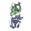
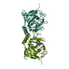
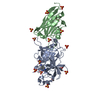
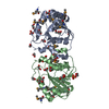
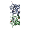
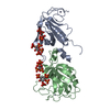
- PDB-1iag: FIRST STRUCTURE OF A SNAKE VENOM METALLOPROTEINASE: A PROTOTYPE F... -
+ Open data

- Basic information
| Entry | Database: PDB / ID: 1iag | ||||||
|---|---|---|---|---|---|---|---|
| Title | FIRST STRUCTURE OF A SNAKE VENOM METALLOPROTEINASE: A PROTOTYPE FOR MATRIX METALLOPROTEINASES(SLASH)COLLAGENASES | ||||||
 Components Components | ADAMALYSIN II Adamalysin Adamalysin | ||||||
 Keywords Keywords | METALLOPROTEASE | ||||||
| Function / homology |  Function and homology informationadamalysin / metalloendopeptidase activity / proteolysis / extracellular region / metal ion binding Function and homology informationadamalysin / metalloendopeptidase activity / proteolysis / extracellular region / metal ion bindingSimilarity search - Function | ||||||
| Biological species |  Crotalus adamanteus (eastern diamondback rattlesnake) Crotalus adamanteus (eastern diamondback rattlesnake) | ||||||
| Method | X-RAY DIFFRACTION / Resolution: 2 Å | ||||||
 Authors Authors | Gomis-Rueth, F.-X. / Bode, W. | ||||||
 Citation Citation | Journal: EMBO J. / Year: 1993 Title: First structure of a snake venom metalloproteinase: a prototype for matrix metalloproteinases/collagenases. Authors: Gomis-Ruth, F.X. / Kress, L.F. / Bode, W. #1: Journal: J.Mol.Biol. / Year: 1994Title: Refined 2.0 Angstroms X-Ray Crystal Structure of the Snake Venom Zinc-Endopeptidase Adamalysin II. Primary and Tertiary Structure Determination, Refinement, Molecular Structure and Comparison ...Title: Refined 2.0 Angstroms X-Ray Crystal Structure of the Snake Venom Zinc-Endopeptidase Adamalysin II. Primary and Tertiary Structure Determination, Refinement, Molecular Structure and Comparison with Astacin, Collagenase and Thermolysin Authors: Gomis-Rueth, F.X. / Kress, L.F. / Kellermann, J. / Mayr, I. / Lee, X. / Huber, R. / Bode, W. #2: Journal: Embo J. / Year: 1994Title: The X-Ray Crystal Structure of the Catalytic Domain of Human Neutrophil Collagenase Inhibited by a Substrate Analogue Reveals the Essentials for Catalysis and Specificity Authors: Bode, W. / Reinemer, P. / Huber, R. / Kleine, T. / Schnierer, S. / Tschesche, H. #3: Journal: FEBS Lett. / Year: 1994Title: Structural Implications for the Role of the N Terminus in the 'Superactivation' of Collagenases. A Crystallographic Study Authors: Reinemer, P. / Grams, F. / Huber, R. / Kleine, T. / Schnierer, S. / Piper, M. / Tschesche, H. / Bode, W. #4: Journal: FEBS Lett. / Year: 1993Title: Astacins, Serralysins, Snake Venom and Matrix Metalloproteinases Exhibit Identical Zinc-Binding Environments (Hexxhxxgxxh and met-Turn) and Topologies and Should be Grouped Into a Common Family, the 'Metzincins' Authors: Bode, W. / Gomis-Rueth, F.-X. / Stoecker, W. #5: Journal: Nature / Year: 1992Title: Structure of Astacin and Implications for Activation of Astacins and Zinc-Ligation of Collagenases Authors: Bode, W. / Gomis-Rueth, F.X. / Huber, R. / Zwilling, R. / Stoecker, W. | ||||||
| History |
|
- Structure visualization
| Structure viewer | Molecule: MolmilJmol/JSmol |
|---|
- Downloads & links
-Download
| PDBx/mmCIF format | 1iag.cif.gz | 53.8 KB | Display | PDBx/mmCIF format |
|---|---|---|---|---|
| PDB format | pdb1iag.ent.gz | 42.3 KB | Display | PDB format |
| PDBx/mmJSON format | 1iag.json.gz | Tree view | PDBx/mmJSON format | |
| Others |  Other downloads Other downloads |
-Validation report
| Arichive directory | https://data.pdbj.org/pub/pdb/validation_reports/ia/1iagftp://data.pdbj.org/pub/pdb/validation_reports/ia/1iag | HTTPS FTP |
|---|
-Related structure data
| Similar structure data |
|---|


-Links
 PDBj
PDBj


- Assembly
| Deposited unit | 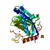
| ||||||||
|---|---|---|---|---|---|---|---|---|---|
| 1 | 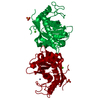
| ||||||||
| Unit cell |
|
-Components
| #1: Protein | Adamalysin Mass: 23211.639 Da / Num. of mol.: 1 Source method: isolated from a genetically manipulated source Source: (gene. exp.) Crotalus adamanteus (eastern diamondback rattlesnake)References: UniProt: P34179, adamalysin |
|---|---|
| #2: Chemical | ChemComp-ZN /   Mass: 65.409 Da / Num. of mol.: 1 / Source method: obtained synthetically / Formula: Zn Mass: 65.409 Da / Num. of mol.: 1 / Source method: obtained synthetically / Formula: Zn |
| #3: Chemical | ChemComp-CA /   Mass: 40.078 Da / Num. of mol.: 1 / Source method: obtained synthetically / Formula: Ca Mass: 40.078 Da / Num. of mol.: 1 / Source method: obtained synthetically / Formula: Ca |
| #4: Chemical | ChemComp-SO4 / Sulfate  Mass: 96.063 Da / Num. of mol.: 1 / Source method: obtained synthetically / Formula: SO4 Mass: 96.063 Da / Num. of mol.: 1 / Source method: obtained synthetically / Formula: SO4 |
| #5: Water | ChemComp-HOH / Water Mass: 18.015 Da / Num. of mol.: 173 / Source method: isolated from a natural source / Formula: H2O Mass: 18.015 Da / Num. of mol.: 173 / Source method: isolated from a natural source / Formula: H2O |
-Experimental details
-Experiment
| Experiment | Method: X-RAY DIFFRACTION |
|---|
- Sample preparation
| Crystal | Density Matthews: 3.25 Å3/Da / Density % sol: 62.11 % |
|---|---|
| Crystal grow | *PLUS pH: 5 / Method: unknown |
| Components of the solutions | *PLUS Conc.: 1.8 M / Common name: ammonium sulfate |
-Data collection
| Radiation | Scattering type: x-ray |
|---|---|
| Radiation wavelength | Relative weight: 1 |
| Reflection | *PLUS Highest resolution: 2 Å / Num. obs: 17021 / % possible obs: 83 % / Num. measured all: 24244 / Rmerge(I) obs: 0.0544 |
| Reflection shell | *PLUS Highest resolution: 2.03 Å / Lowest resolution: 2.09 Å / % possible obs: 42 % |
- Processing
| Software |
| ||||||||||||||||||||||||||||||||||||||||||||||||||||||||||||
|---|---|---|---|---|---|---|---|---|---|---|---|---|---|---|---|---|---|---|---|---|---|---|---|---|---|---|---|---|---|---|---|---|---|---|---|---|---|---|---|---|---|---|---|---|---|---|---|---|---|---|---|---|---|---|---|---|---|---|---|---|---|
| Refinement | Resolution: 2→8 Å / Rfactor Rwork: 0.172 / Rfactor obs: 0.172 Details: 158 OF THE 203 RESIDUES HAVE BEEN CHECKED BY PEPTIDE SEQUENCING. THE REMAINING 45 AMINO ACID RESIDUES HAVE BEEN IDENTIFIED ONLY FROM THEIR DENSITY (FOR DISCUSSION, SEE GOMIS-RUETH ET AL. ...Details: 158 OF THE 203 RESIDUES HAVE BEEN CHECKED BY PEPTIDE SEQUENCING. THE REMAINING 45 AMINO ACID RESIDUES HAVE BEEN IDENTIFIED ONLY FROM THEIR DENSITY (FOR DISCUSSION, SEE GOMIS-RUETH ET AL. (1994) J.MOL.BIOL, 239, 513-544, AND FIGURES 1 AND 2. | ||||||||||||||||||||||||||||||||||||||||||||||||||||||||||||
| Refinement step | Cycle: LAST / Resolution: 2→8 Å
| ||||||||||||||||||||||||||||||||||||||||||||||||||||||||||||
| Refine LS restraints |
| ||||||||||||||||||||||||||||||||||||||||||||||||||||||||||||
| Software | *PLUS Name: X-PLOR / Classification: refinement | ||||||||||||||||||||||||||||||||||||||||||||||||||||||||||||
| Refinement | *PLUS Num. reflection obs: 16776 / Rfactor obs: 0.172 | ||||||||||||||||||||||||||||||||||||||||||||||||||||||||||||
| Solvent computation | *PLUS | ||||||||||||||||||||||||||||||||||||||||||||||||||||||||||||
| Displacement parameters | *PLUS | ||||||||||||||||||||||||||||||||||||||||||||||||||||||||||||
| Refine LS restraints | *PLUS Type: x_angle_d / Dev ideal: 1.9 |
