 Movie
Movie Controller


+ Open data

- Basic information
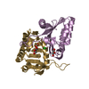
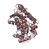
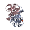
| Entry | Database: PDB / ID: 1i3c | ||||||
|---|---|---|---|---|---|---|---|
| Title | RESPONSE REGULATOR FOR CYANOBACTERIAL PHYTOCHROME, RCP1 | ||||||
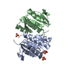
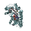
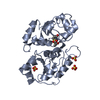
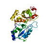
 Components Components | RESPONSE REGULATOR RCP1 | ||||||
 Keywords Keywords | SIGNALING PROTEIN / Response regulator / Rcp1 / Phytochrome | ||||||
| Function / homology |  Function and homology information Function and homology information | ||||||
| Biological species |  | ||||||
| Method |  X-RAY DIFFRACTION / SYNCHROTRON / MAD / Resolution: 1.9 Å X-RAY DIFFRACTION / SYNCHROTRON / MAD / Resolution: 1.9 Å | ||||||
 Authors Authors | Im, Y.J. / Rho, S.-H. / Park, C.-M. / Yang, S.-S. / Kang, J.-G. / Lee, J.Y. / Song, P.-S. / Eom, S.H. | ||||||
 Citation Citation | Journal: Protein Sci. / Year: 2002 Title: Crystal structure of a cyanobacterial phytochrome response regulator. Authors: Im, Y.J. / Rho, S.H. / Park, C.M. / Yang, S.S. / Kang, J.G. / Lee, J.Y. / Song, P.S. / Eom, S.H. | ||||||
| History |
|
- Structure visualization
| Structure viewer | Molecule: MolmilJmol/JSmol |
|---|
- Downloads & links
-Download
| PDBx/mmCIF format | 1i3c.cif.gz | 75.1 KB | Display | PDBx/mmCIF format |
|---|---|---|---|---|
| PDB format | pdb1i3c.ent.gz | 55.5 KB | Display | PDB format |
| PDBx/mmJSON format | 1i3c.json.gz | Tree view | PDBx/mmJSON format | |
| Others |  Other downloads Other downloads |
-Validation report
| Arichive directory | https://data.pdbj.org/pub/pdb/validation_reports/i3/1i3cftp://data.pdbj.org/pub/pdb/validation_reports/i3/1i3c | HTTPS FTP |
|---|
-Related structure data


-Links
 PDBj
PDBj

- Assembly
| Deposited unit | 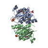
| ||||||||
|---|---|---|---|---|---|---|---|---|---|
| 1 |
| ||||||||
| Unit cell |
|
-Components
| #1: Protein | Mass: 16922.898 Da / Num. of mol.: 2 Source method: isolated from a genetically manipulated source Source: (gene. exp.) #2: Chemical | ChemComp-SO4 /   Mass: 96.063 Da / Num. of mol.: 5 / Source method: obtained synthetically / Formula: SO4 Mass: 96.063 Da / Num. of mol.: 5 / Source method: obtained synthetically / Formula: SO4#3: Water | ChemComp-HOH / |  Mass: 18.015 Da / Num. of mol.: 269 / Source method: isolated from a natural source / Formula: H2O Mass: 18.015 Da / Num. of mol.: 269 / Source method: isolated from a natural source / Formula: H2OHas protein modification | Y | |
|---|
-Experimental details
-Experiment
| Experiment | Method: X-RAY DIFFRACTION / Number of used crystals: 1 |
|---|
- Sample preparation
| Crystal | Density Matthews: 1.95 Å3/Da / Density % sol: 36.8 % | ||||||||||||||||||
|---|---|---|---|---|---|---|---|---|---|---|---|---|---|---|---|---|---|---|---|
| Crystal grow | Temperature: 294 K / Method: vapor diffusion, hanging drop / pH: 8.5 Details: Ammonium Sulfate, Tris, pH 8.5, VAPOR DIFFUSION, HANGING DROP, temperature 294K | ||||||||||||||||||
| Crystal grow | *PLUS Temperature: 293-295 K | ||||||||||||||||||
| Components of the solutions | *PLUS
|
-Data collection
| Diffraction |
| ||||||||||||||||||||
|---|---|---|---|---|---|---|---|---|---|---|---|---|---|---|---|---|---|---|---|---|---|
| Diffraction source |
| ||||||||||||||||||||
| Detector | Type: ADSC QUANTUM 4 / Detector: CCD / Date: Jul 15, 2000 / Details: Quartz crystal | ||||||||||||||||||||
| Radiation | Monochromator: Quartz crystal / Protocol: MAD / Monochromatic (M) / Laue (L): M / Scattering type: x-ray | ||||||||||||||||||||
| Radiation wavelength |
| ||||||||||||||||||||
| Reflection | Resolution: 1.9→14.97 Å / Num. all: 20164 / Num. obs: 83143 / % possible obs: 98.5 % / Observed criterion σ(F): 0 / Observed criterion σ(I): 4 / Redundancy: 4 % / Biso Wilson estimate: 10.1 Å2 / Limit h max: 20 / Limit h min: -21 / Limit k max: 40 / Limit k min: -21 / Limit l max: 22 / Limit l min: 0 / Observed criterion F max: 892718.68 / Observed criterion F min: 7.4 / Rmerge(I) obs: 0.046 / Net I/σ(I): 12.8 | ||||||||||||||||||||
| Reflection shell | Resolution: 1.9→1.99 Å / Redundancy: 4 % / Rmerge(I) obs: 0.155 / % possible all: 98.5 | ||||||||||||||||||||
| Reflection | *PLUS Highest resolution: 1.9 Å / Lowest resolution: 15 Å / Num. obs: 20025 / Num. measured all: 83143 | ||||||||||||||||||||
| Reflection shell | *PLUS % possible obs: 98.5 % |

- Processing
| Software |
| ||||||||||||||||||||||||||||||||||||||||||||||||||||||||||||||||||||||||||||||||||||||||||
|---|---|---|---|---|---|---|---|---|---|---|---|---|---|---|---|---|---|---|---|---|---|---|---|---|---|---|---|---|---|---|---|---|---|---|---|---|---|---|---|---|---|---|---|---|---|---|---|---|---|---|---|---|---|---|---|---|---|---|---|---|---|---|---|---|---|---|---|---|---|---|---|---|---|---|---|---|---|---|---|---|---|---|---|---|---|---|---|---|---|---|---|
| Refinement | Method to determine structure: MAD / Resolution: 1.9→14.97 Å / Rfactor Rfree error: 0.005 / Occupancy max: 1 / Occupancy min: 1 / Isotropic thermal model: Isotropic / Cross valid method: THROUGHOUT / σ(F): 2 / σ(I): 4 / Stereochemistry target values: Engh & Huber
| ||||||||||||||||||||||||||||||||||||||||||||||||||||||||||||||||||||||||||||||||||||||||||
| Solvent computation | Solvent model: CNS bulk solvent model used / Bsol: 42.347 Å2 / ksol: 0.391785 e/Å3 | ||||||||||||||||||||||||||||||||||||||||||||||||||||||||||||||||||||||||||||||||||||||||||
| Displacement parameters | Biso max: 56.2 Å2 / Biso mean: 14.51 Å2 / Biso min: 1.48 Å2
| ||||||||||||||||||||||||||||||||||||||||||||||||||||||||||||||||||||||||||||||||||||||||||
| Refine analyze |
| ||||||||||||||||||||||||||||||||||||||||||||||||||||||||||||||||||||||||||||||||||||||||||
| Refinement step | Cycle: LAST / Resolution: 1.9→14.97 Å
| ||||||||||||||||||||||||||||||||||||||||||||||||||||||||||||||||||||||||||||||||||||||||||
| Refine LS restraints |
| ||||||||||||||||||||||||||||||||||||||||||||||||||||||||||||||||||||||||||||||||||||||||||
| LS refinement shell | Refine-ID: X-RAY DIFFRACTION
| ||||||||||||||||||||||||||||||||||||||||||||||||||||||||||||||||||||||||||||||||||||||||||
| Xplor file |
| ||||||||||||||||||||||||||||||||||||||||||||||||||||||||||||||||||||||||||||||||||||||||||
| Refinement | *PLUS Highest resolution: 1.9 Å / Lowest resolution: 15 Å / σ(F): 2 / % reflection Rfree: 9.9 % / Rfactor obs: 0.188 | ||||||||||||||||||||||||||||||||||||||||||||||||||||||||||||||||||||||||||||||||||||||||||
| Solvent computation | *PLUS | ||||||||||||||||||||||||||||||||||||||||||||||||||||||||||||||||||||||||||||||||||||||||||
| Displacement parameters | *PLUS Biso mean: 14.5 Å2 | ||||||||||||||||||||||||||||||||||||||||||||||||||||||||||||||||||||||||||||||||||||||||||
| LS refinement shell | *PLUS Rfactor Rfree: 0.219 / % reflection Rfree: 9.4 % / Rfactor Rwork: 0.221 |
