 Movie
Movie Controller


+ Open data

- Basic information
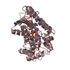
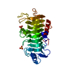
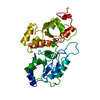
| Entry | Database: PDB / ID: 1dp2 | ||||||
|---|---|---|---|---|---|---|---|
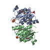
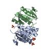
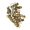
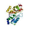
| Title | CRYSTAL STRUCTURE OF THE COMPLEX BETWEEN RHODANESE AND LIPOATE | ||||||
 Components Components | RHODANESE | ||||||
 Keywords Keywords | TRANSFERASE / Rhodanese / liopate / sulfurtransferase | ||||||
| Function / homology |  Function and homology information Function and homology informationrRNA transport / 3-mercaptopyruvate sulfurtransferase activity / thiosulfate sulfurtransferase / thiosulfate-cyanide sulfurtransferase activity / rRNA import into mitochondrion / 5S rRNA binding / mitochondrial matrix / mitochondrion Similarity search - Function | ||||||
| Biological species |  | ||||||
| Method |  X-RAY DIFFRACTION / Resolution: 2.01 Å X-RAY DIFFRACTION / Resolution: 2.01 Å | ||||||
 Authors Authors | Zanotti, G. / Cianci, M. | ||||||
 Citation Citation | Journal: Biochim.Biophys.Acta / Year: 2000 Title: Specific interaction of lipoate at the active site of rhodanese. Authors: Cianci, M. / Gliubich, F. / Zanotti, G. / Berni, R. #1: Journal: J.Mol.Biol. / Year: 1979Title: THE STRUCTURE OF BOVINE LIVER RHODANESE. II. THE ACTIVE SITE IN THE SULFUR-SUBSTITUTED AND THE SULFUR-FREE ENZYME Authors: Ploegman, J.H. / Drenth, G. / Kalk, K.H. / Hol, W.G.J. #2: Journal: Acta Crystallogr.,Sect.D / Year: 1998Title: ACTIVE SITE STRUCTURAL FEATURES FOR CHEMICALLY MODIFIED FORMS OF RHODANESE Authors: Gliubich, F. / Berni, R. / Colapietro, M. / Barba, L. / Zanotti, G. | ||||||
| History |
|
- Structure visualization
| Structure viewer | Molecule: MolmilJmol/JSmol |
|---|
- Downloads & links
-Download
| PDBx/mmCIF format | 1dp2.cif.gz | 72.7 KB | Display | PDBx/mmCIF format |
|---|---|---|---|---|
| PDB format | pdb1dp2.ent.gz | 54 KB | Display | PDB format |
| PDBx/mmJSON format | 1dp2.json.gz | Tree view | PDBx/mmJSON format | |
| Others |  Other downloads Other downloads |
-Validation report
| Arichive directory | https://data.pdbj.org/pub/pdb/validation_reports/dp/1dp2ftp://data.pdbj.org/pub/pdb/validation_reports/dp/1dp2 | HTTPS FTP |
|---|
-Related structure data
| Related structure data | |
|---|---|
| Similar structure data |


-Links
 PDBj
PDBj- Assembly
| Deposited unit | 
| ||||||||
|---|---|---|---|---|---|---|---|---|---|
| 1 |
| ||||||||
| Unit cell |
|
-Components
| #1: Protein | Mass: 32983.359 Da / Num. of mol.: 1 / Source method: isolated from a natural source / Source: (natural) |
|---|---|
| #2: Chemical | ChemComp-LPB / 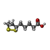  Mass: 206.326 Da / Num. of mol.: 1 / Source method: obtained synthetically / Formula: C8H14O2S2 Mass: 206.326 Da / Num. of mol.: 1 / Source method: obtained synthetically / Formula: C8H14O2S2 |
| #3: Water | ChemComp-HOH /  Mass: 18.015 Da / Num. of mol.: 112 / Source method: isolated from a natural source / Formula: H2O Mass: 18.015 Da / Num. of mol.: 112 / Source method: isolated from a natural source / Formula: H2O |
| Has protein modification | Y |
-Experimental details
-Experiment
| Experiment | Method: X-RAY DIFFRACTION / Number of used crystals: 2 |
|---|
- Sample preparation
| Crystal | Density Matthews: 2.37 Å3/Da / Density % sol: 48.09 % | |||||||||||||||
|---|---|---|---|---|---|---|---|---|---|---|---|---|---|---|---|---|
| Crystal grow | Temperature: 293 K / Method: vapor diffusion, sitting drop / pH: 7 Details: Crystals were obtained from ammonium sulfate. After soaking with 28% PEG 6000, 40 mM phosphate buffer, pH=7, 4 mM DL-lipoate was added , VAPOR DIFFUSION, SITTING DROP, temperature 20K, temperature 293K | |||||||||||||||
| Crystal grow | *PLUS pH: 7.4 / Details: Gliubich, F., (1996), J. Biol. Chem., 271, 21054. | |||||||||||||||
| Components of the solutions | *PLUS
|
-Data collection
| Diffraction | Mean temperature: 300 K |
|---|---|
| Diffraction source | Source: ROTATING ANODE / Type: SIEMENS / Wavelength: 1.5418 |
| Detector | Type: SIEMENS HI-STAR / Detector: AREA DETECTOR / Date: Jun 11, 1998 |
| Radiation | Protocol: SINGLE WAVELENGTH / Monochromatic (M) / Laue (L): M / Scattering type: x-ray |
| Radiation wavelength | Wavelength: 1.5418 Å / Relative weight: 1 |
| Reflection | Resolution: 2.01→55 Å / Num. all: 18241 / Num. obs: 18062 / % possible obs: 85.6 % / Observed criterion σ(F): 0 / Observed criterion σ(I): 0 / Redundancy: 2.3 % / Rmerge(I) obs: 0.043 / Net I/σ(I): 14 |
| Reflection shell | Resolution: 2.01→2.2 Å / Redundancy: 1.8 % / Rmerge(I) obs: 0.134 / Num. unique all: 3174 / % possible all: 68 |
| Reflection | *PLUS Num. obs: 18241 / Num. measured all: 42252 |
| Reflection shell | *PLUS % possible obs: 68 % |
- Processing
| Software |
| ||||||||||||||||||||
|---|---|---|---|---|---|---|---|---|---|---|---|---|---|---|---|---|---|---|---|---|---|
| Refinement | Resolution: 2.01→55 Å / σ(F): 0 / σ(I): 0 / Stereochemistry target values: Param19X.pro (XPLOR)
| ||||||||||||||||||||
| Refinement step | Cycle: LAST / Resolution: 2.01→55 Å
| ||||||||||||||||||||
| Refine LS restraints |
| ||||||||||||||||||||
| Software | *PLUS Name: X-PLOR / Version: 3.1 / Classification: refinement | ||||||||||||||||||||
| Refine LS restraints | *PLUS Type: x_bond_d / Dev ideal: 0.01 |
