 Movie
Movie Controller

[English] 日本語
 Yorodumi
- PDB-1hyb: CRYSTAL STRUCTURE OF AN ACTIVE SITE MUTANT OF METHANOBACTERIUM TH... -
+ Open data

- Basic information
| Entry | Database: PDB / ID: 1hyb | ||||||
|---|---|---|---|---|---|---|---|
| Title | CRYSTAL STRUCTURE OF AN ACTIVE SITE MUTANT OF METHANOBACTERIUM THERMOAUTOTROPHICUM NICOTINAMIDE MONONUCLEOTIDE ADENYLYLTRANSFERASE | ||||||
 Components Components | NICOTINAMIDE MONONUCLEOTIDE ADENYLYLTRANSFERASE | ||||||
 Keywords Keywords |  TRANSFERASE / dinucleotide binding fold / active site mutant TRANSFERASE / dinucleotide binding fold / active site mutant | ||||||
| Function / homology |  Function and homology informationnicotinamide-nucleotide adenylyltransferase / nicotinamide-nucleotide adenylyltransferase activity / NAD biosynthetic process / ATP binding / cytoplasm Function and homology informationnicotinamide-nucleotide adenylyltransferase / nicotinamide-nucleotide adenylyltransferase activity / NAD biosynthetic process / ATP binding / cytoplasmSimilarity search - Function | ||||||
| Biological species |   Methanothermobacter thermautotrophicus (archaea) Methanothermobacter thermautotrophicus (archaea) | ||||||
| Method | X-RAY DIFFRACTION / SYNCHROTRON / isomorphous to native / Resolution: 2 Å | ||||||
 Authors Authors | Saridakis, V. / Christendat, D. / Kimber, M.S. / Edwards, A.M. / Pai, E.F. | ||||||
 Citation Citation | Journal: J.Biol.Chem. / Year: 2001 Title: Insights into ligand binding and catalysis of a central step in NAD+ synthesis: structures of Methanobacterium thermoautotrophicum NMN adenylyltransferase complexes. Authors: Saridakis, V. / Christendat, D. / Kimber, M.S. / Dharamsi, A. / Edwards, A.M. / Pai, E.F. | ||||||
| History |
|
- Structure visualization
| Structure viewer | Molecule: MolmilJmol/JSmol |
|---|
- Downloads & links
-Download
| PDBx/mmCIF format | 1hyb.cif.gz | 48.2 KB | Display | PDBx/mmCIF format |
|---|---|---|---|---|
| PDB format | pdb1hyb.ent.gz | 33.1 KB | Display | PDB format |
| PDBx/mmJSON format | 1hyb.json.gz | Tree view | PDBx/mmJSON format | |
| Others |  Other downloads Other downloads |
-Validation report
| Arichive directory | https://data.pdbj.org/pub/pdb/validation_reports/hy/1hybftp://data.pdbj.org/pub/pdb/validation_reports/hy/1hyb | HTTPS FTP |
|---|
-Related structure data
| Related structure data |  1ej2SC S: Starting model for refinement C: citing same article ( |
|---|---|
| Similar structure data |


-Links
 PDBj
PDBj- Assembly
| Deposited unit | 
| ||||||||
|---|---|---|---|---|---|---|---|---|---|
| 1 | x 6
| ||||||||
| Unit cell |
| ||||||||
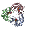
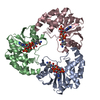
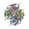
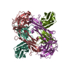
| Details | The biological assembly is a hexamer constructed from chain A and five symmetry partners generated by crystallographic symmetry. |
-Components
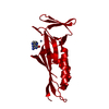
| #1: Protein | Mass: 20529.725 Da / Num. of mol.: 1 / Mutation: H19A Source method: isolated from a genetically manipulated source Source: (gene. exp.) Methanothermobacter thermautotrophicus (archaea)Plasmid: PET15B / Species (production host): Escherichia coli / Production host:  Escherichia coli BL21(DE3) (bacteria) / Strain (production host): BL21 (DE3) Escherichia coli BL21(DE3) (bacteria) / Strain (production host): BL21 (DE3)References: UniProt: O26253, nicotinamide-nucleotide adenylyltransferase |
|---|---|
| #2: Chemical | ChemComp-SO4 / Sulfate  Mass: 96.063 Da / Num. of mol.: 1 / Source method: obtained synthetically / Formula: SO4 Mass: 96.063 Da / Num. of mol.: 1 / Source method: obtained synthetically / Formula: SO4 |
| #3: Chemical | ChemComp-NMN / Nicotinamide mononucleotide  Mass: 335.227 Da / Num. of mol.: 1 / Source method: obtained synthetically / Formula: C11H16N2O8P Mass: 335.227 Da / Num. of mol.: 1 / Source method: obtained synthetically / Formula: C11H16N2O8P |
| #4: Water | ChemComp-HOH / Water Mass: 18.015 Da / Num. of mol.: 70 / Source method: isolated from a natural source / Formula: H2O Mass: 18.015 Da / Num. of mol.: 70 / Source method: isolated from a natural source / Formula: H2O |
-Experimental details
-Experiment
| Experiment | Method: X-RAY DIFFRACTION / Number of used crystals: 2 |
|---|
- Sample preparation
| Crystal | Density Matthews: 3.09 Å3/Da / Density % sol: 60.13 % | ||||||||||||||||||||||||
|---|---|---|---|---|---|---|---|---|---|---|---|---|---|---|---|---|---|---|---|---|---|---|---|---|---|
| Crystal grow | Temperature: 298 K / Method: vapor diffusion, hanging drop / pH: 7.5 Details: 1.6 M LiSO4, 100 mM HEPES, pH 7.5, VAPOR DIFFUSION, HANGING DROP, temperature 298K | ||||||||||||||||||||||||
| Crystal grow | *PLUS Temperature: 20 ℃ | ||||||||||||||||||||||||
| Components of the solutions | *PLUS
|
-Data collection
| Diffraction | Mean temperature: 100 K |
|---|---|
| Diffraction source | Source: SYNCHROTRON / Site: APS  / Beamline: 14-BM-C / Wavelength: 1 Å / Beamline: 14-BM-C / Wavelength: 1 Å |
| Detector | Type: ADSC QUANTUM 4 / Detector: CCD / Date: Nov 28, 2000 |
| Radiation | Protocol: SINGLE WAVELENGTH / Monochromatic (M) / Laue (L): M / Scattering type: x-ray |
| Radiation wavelength | Wavelength: 1 Å / Relative weight: 1 |
| Reflection | Resolution: 2→30 Å / Num. all: 253617 / Num. obs: 17892 / % possible obs: 98.1 % / Observed criterion σ(F): 16525 / Observed criterion σ(I): 486 / Redundancy: 12 % / Biso Wilson estimate: 23.7 Å2 / Rmerge(I) obs: 0.073 / Net I/σ(I): 34 |
| Reflection shell | Resolution: 2→30 Å / Redundancy: 10 % / Rmerge(I) obs: 0.337 / Mean I/σ(I) obs: 7 / Num. unique all: 1710 / % possible all: 98.1 |
| Reflection shell | *PLUS % possible obs: 98.1 % |
- Processing
| Software |
| ||||||||||||||||||||||||||||||||||||
|---|---|---|---|---|---|---|---|---|---|---|---|---|---|---|---|---|---|---|---|---|---|---|---|---|---|---|---|---|---|---|---|---|---|---|---|---|---|
| Refinement | Method to determine structure: isomorphous to native Starting model: pdb 1ej2 Resolution: 2→14.96 Å / Rfactor Rfree error: 0.007 / Data cutoff high absF: 929092.67 / Data cutoff low absF: 0 / Isotropic thermal model: RESTRAINED / Cross valid method: THROUGHOUT / Stereochemistry target values: CNS 0.9 / Details: simulated annealing
| ||||||||||||||||||||||||||||||||||||
| Solvent computation | Solvent model: FLAT MODEL / Bsol: 55.48 Å2 / ksol: 0.409 e/Å3 | ||||||||||||||||||||||||||||||||||||
| Displacement parameters | Biso mean: 37.4 Å2
| ||||||||||||||||||||||||||||||||||||
| Refine analyze |
| ||||||||||||||||||||||||||||||||||||
| Refinement step | Cycle: LAST / Resolution: 2→14.96 Å
| ||||||||||||||||||||||||||||||||||||
| Refine LS restraints |
| ||||||||||||||||||||||||||||||||||||
| LS refinement shell | Resolution: 2→2.12 Å / Rfactor Rfree error: 0.019 / Total num. of bins used: 6
| ||||||||||||||||||||||||||||||||||||
| Xplor file |
| ||||||||||||||||||||||||||||||||||||
| Software | *PLUS Name: CNS / Version: 0.9 / Classification: refinement | ||||||||||||||||||||||||||||||||||||
| Refine LS restraints | *PLUS
|
